W3 CH 3 - Neurophysiology
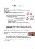
Glia and Neurons
Glia
Glia - Cells in the nervous system that support the activities of neurons.
Neuron - A cell of the nervous system that is specialized for information processing and
communication.
Macroglia - Large glial cells, including astrocytes, ependymal cells,
oligodendrocytes, and Schwann cells.
- Astrocyte - A large, star shaped glial cell of the central nervous
system (CNS), responsible for structural support, isolation of the
synapse, control of the extracellular chemical environment at the
synapse, and possibly communication.
- The close association of astrocytes with the capillary cells
allows these glia to transfer glucose and other nutrients to
the neurons. Because of their ability to contact both blood
vessels and synapses, or points of communication
between two cells, astrocytes regulate local blood flow
based on synaptic activity.
- Synapse - The junction between two neurons at which information is
transferred from one to another.
- Blood-brain barrier - An impediment to the transfer of molecules from the
circulation into the brain formed by the astrocytes.
- Prevents most toxins circulating in the blood from entering the brain.
- Ependymal cells - Glial cells lining the ventricles and central canal of the spinal cord.
- Oligodendrocyte - A glial cell that forms the myelin on central nervous system (CNS)
axons.
- Schwann cell - A glial cell that forms the myelin on axons in the peripheral nervous
system (PNS).
Microglia - Tiny, mobile glial cells that migrate to areas of damage and digest debris.
EPENDYMAL CELLS -> Glial cells lining the ventricles and central canal of the
spinal cord.
- Ependymal cells feature fine hair-like cilia that project into a ventricle or
the central canal and move cerebrospinal fluid (CSF) with a whip-like
motion.
- The cilia also absorb some CSF, allowing the ependymal cells to monitor
the quality of the CSF and to supply underlying brain cells with proteins from the CSF.
OLIGODENDROCYTES AND SCHWANN CELLS -> Oligodendrocytes and Schwann cells
provide the myelin covering that insulates some nerve fibers or axons (Oligo for CNS and
Schwann for PNS)
, - Oligodendrocytes and Schwann cells can actually
communicate with their nearby axons by releasing little
packets of material (ventricles) known as exosomes ->
remove debris from a cell and also appear to deliver
substances, including genetic material, to other cells.
MICROGLIA -> Tiny, mobile glial cells that migrate to areas of
damage and digest debris.
- Uncontrolled activation of microglia, though, can damage
the brain. Microglia have been observed digesting healthy
cells located next to damaged cells (Kim & Joh, 2006).
Inflammation caused by microglia activation is under
investigation as a contributor to neurodegenerative diseases,
including Alzheimer's disease, Parkinson's disease, and multiple sclerosis.
- Microglia play a role in the removal of less active synapses, which is an important part of
the wiring of the developing brain.
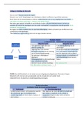
, The Structure of Neurons
- All animal cells, including neurons, have membranes, nuclei, and small internal
structures known as organelles. Many of these structures are found within the main
mass of the neuron, known as the cell body/soma.
- Neurons differ structurally from other cells in that they have specialized branches
extending from the cell body, known as the axons and dendrites, which they use to
communicate with other cells.
NEURAL MEMBRANES
- Form a boundary between the cell and its external environment. The neural membrane
must separate the intracellular fluid or cytoplasm of the cell's interior from the
extracellular fluid surrounding the neuron.
- Made up of a double layer of phospholipids, fatty molecules that contain phosphate.
Because they are fats, phospholipids do not dissolve in water.
- Suspended within this phospholipid membrane are a number of important protein
structures that control its permeability [movement of substances across the cell
membrane].
- There are two primary types of protein structures of
interest in our discussion of neural function: ion
channels and ion pumps. These structures provide
pores, or channels, through which specific ions, or
electrically charged particles, can move into or out of
the neuron. Ion channels allow ions to move passively,
without the expenditure of energy, whereas ion pumps
require energy.
- Voltage-dependent channel -An ion channel that
opens or closes in response to the local electrical
environment.
- Ligand-gated channel - An ion channel in the neural
membrane that responds to chemical messengers.
Glia and Neurons
Glia
Glia - Cells in the nervous system that support the activities of neurons.
Neuron - A cell of the nervous system that is specialized for information processing and
communication.
Macroglia - Large glial cells, including astrocytes, ependymal cells,
oligodendrocytes, and Schwann cells.
- Astrocyte - A large, star shaped glial cell of the central nervous
system (CNS), responsible for structural support, isolation of the
synapse, control of the extracellular chemical environment at the
synapse, and possibly communication.
- The close association of astrocytes with the capillary cells
allows these glia to transfer glucose and other nutrients to
the neurons. Because of their ability to contact both blood
vessels and synapses, or points of communication
between two cells, astrocytes regulate local blood flow
based on synaptic activity.
- Synapse - The junction between two neurons at which information is
transferred from one to another.
- Blood-brain barrier - An impediment to the transfer of molecules from the
circulation into the brain formed by the astrocytes.
- Prevents most toxins circulating in the blood from entering the brain.
- Ependymal cells - Glial cells lining the ventricles and central canal of the spinal cord.
- Oligodendrocyte - A glial cell that forms the myelin on central nervous system (CNS)
axons.
- Schwann cell - A glial cell that forms the myelin on axons in the peripheral nervous
system (PNS).
Microglia - Tiny, mobile glial cells that migrate to areas of damage and digest debris.
EPENDYMAL CELLS -> Glial cells lining the ventricles and central canal of the
spinal cord.
- Ependymal cells feature fine hair-like cilia that project into a ventricle or
the central canal and move cerebrospinal fluid (CSF) with a whip-like
motion.
- The cilia also absorb some CSF, allowing the ependymal cells to monitor
the quality of the CSF and to supply underlying brain cells with proteins from the CSF.
OLIGODENDROCYTES AND SCHWANN CELLS -> Oligodendrocytes and Schwann cells
provide the myelin covering that insulates some nerve fibers or axons (Oligo for CNS and
Schwann for PNS)
, - Oligodendrocytes and Schwann cells can actually
communicate with their nearby axons by releasing little
packets of material (ventricles) known as exosomes ->
remove debris from a cell and also appear to deliver
substances, including genetic material, to other cells.
MICROGLIA -> Tiny, mobile glial cells that migrate to areas of
damage and digest debris.
- Uncontrolled activation of microglia, though, can damage
the brain. Microglia have been observed digesting healthy
cells located next to damaged cells (Kim & Joh, 2006).
Inflammation caused by microglia activation is under
investigation as a contributor to neurodegenerative diseases,
including Alzheimer's disease, Parkinson's disease, and multiple sclerosis.
- Microglia play a role in the removal of less active synapses, which is an important part of
the wiring of the developing brain.
, The Structure of Neurons
- All animal cells, including neurons, have membranes, nuclei, and small internal
structures known as organelles. Many of these structures are found within the main
mass of the neuron, known as the cell body/soma.
- Neurons differ structurally from other cells in that they have specialized branches
extending from the cell body, known as the axons and dendrites, which they use to
communicate with other cells.
NEURAL MEMBRANES
- Form a boundary between the cell and its external environment. The neural membrane
must separate the intracellular fluid or cytoplasm of the cell's interior from the
extracellular fluid surrounding the neuron.
- Made up of a double layer of phospholipids, fatty molecules that contain phosphate.
Because they are fats, phospholipids do not dissolve in water.
- Suspended within this phospholipid membrane are a number of important protein
structures that control its permeability [movement of substances across the cell
membrane].
- There are two primary types of protein structures of
interest in our discussion of neural function: ion
channels and ion pumps. These structures provide
pores, or channels, through which specific ions, or
electrically charged particles, can move into or out of
the neuron. Ion channels allow ions to move passively,
without the expenditure of energy, whereas ion pumps
require energy.
- Voltage-dependent channel -An ion channel that
opens or closes in response to the local electrical
environment.
- Ligand-gated channel - An ion channel in the neural
membrane that responds to chemical messengers.