Module 2 Revision
2.1 MICROSCOPES
MAGNIFICATION - number of times larger an image appears, compared to the size of the object. microscopes
produce linear magnification (if specimen magnified x100, the width and length are 100 times greater).
MAGNIFICATION = OBJECTIVE LENS X EYEPIECE LENS
MAGNIFICATION FACTOR = IMAGE SIZE / ACTUAL OBJECT SIZE
RESOLUTION - clarity of an image; the higher the resolution, the clearer the image.
PHOTOMICROGRAPH - photograph of an image seen using an optical microscope.
ELECTRON MICROGRAPH - photograph of an image seen using an electron microscope.
OPTICAL MICROSCOPE - uses visible light (part of electromagnetic spectrum, with wavelength 400-700nm).
- minimum distance between two objects seen as separate entities = 200nm (half of minimum wavelength of
illuminating source).
- ribosomes cannot be seen.
- cheap; easy to use; portable; can study whole living organisms.
LASER SCANNING MICROSCOPE - also known as CONFOCAL microscope.
- use laser light to scan an object point by point.
- assemble with the computer the pixel information into an image.
- high resolution and high contrast images.
- depth selectivity - can focus on structures at different depths within specimens. used to clearly observe whole
living specimens.
- used in medicine & biological research - e.g. observe fungal filaments in cornea of eye (fungal cornea
infection), to diagnose earlier and get more effective treatment.
ELECTRON MICROSCOPE - use beams of fast electrons with wavelength 0.004nm (greater resolution than optical
microscopes).
- electrons fired from a cathode, and focused by magnets (not glass lenses) onto screen / photographic plate.
- large; expensive; need a great amount of skill and training to use; dead specimens; hazardous metallic salt
stains (staining).
TRANSMISSION ELECTRON MICROSCOPE - specimen chemically fixed by dehydration and staining.
- a beam of electrons passed through the specimen, which is stained with metal salts. some electrons pass
through and are focused on the screen / photographic plate.
- electrons form a 2D grey-scale image (electron micrograph).
- maximum magnification = x2,000,000.
SCANNING ELECTRON MICROSCOPE - secondary electrons are ‘bounced off’ the specimen’s surface, and
focussed onto a screen.
- 3D grey-scale image. computer software programmes can add false colour.
- magnification ranges from x15 to x200,000.
- specimen placed in vacuum, and coated with fine metal film.
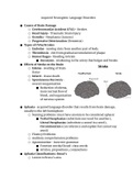
2.1 SLIDES AND MICROGRAPHS
STAINS - coloured chemicals that bind to molecules in/on a specimen, to make it easy to see. many biological
structures are colourless and transparent, so require staining.
- methylene blue: all-purpose stain.
- acetic orcein: binds to DNA. stains chromosomes dark red.
- eosin: stains cytoplasm
- Sudan-red: stains lipids.
- iodine (in potassium iodide solution): stains cellulose in plant cell walls yellow. stains starch granules
blue/black (violet under microscope).
DIFFERENTIAL STAINING - some stains bind to specific cell structures, staining each differently so the structures
can be easily identified.
PREPARING SPECIMENS - dehydrating of specimens; embedding in wax to prevent distortion during slicing; using a
special instrument to make thin sections (sectioning); staining and mounting in a special chemical to preserve them.
2.1 MICROSCOPES
MAGNIFICATION - number of times larger an image appears, compared to the size of the object. microscopes
produce linear magnification (if specimen magnified x100, the width and length are 100 times greater).
MAGNIFICATION = OBJECTIVE LENS X EYEPIECE LENS
MAGNIFICATION FACTOR = IMAGE SIZE / ACTUAL OBJECT SIZE
RESOLUTION - clarity of an image; the higher the resolution, the clearer the image.
PHOTOMICROGRAPH - photograph of an image seen using an optical microscope.
ELECTRON MICROGRAPH - photograph of an image seen using an electron microscope.
OPTICAL MICROSCOPE - uses visible light (part of electromagnetic spectrum, with wavelength 400-700nm).
- minimum distance between two objects seen as separate entities = 200nm (half of minimum wavelength of
illuminating source).
- ribosomes cannot be seen.
- cheap; easy to use; portable; can study whole living organisms.
LASER SCANNING MICROSCOPE - also known as CONFOCAL microscope.
- use laser light to scan an object point by point.
- assemble with the computer the pixel information into an image.
- high resolution and high contrast images.
- depth selectivity - can focus on structures at different depths within specimens. used to clearly observe whole
living specimens.
- used in medicine & biological research - e.g. observe fungal filaments in cornea of eye (fungal cornea
infection), to diagnose earlier and get more effective treatment.
ELECTRON MICROSCOPE - use beams of fast electrons with wavelength 0.004nm (greater resolution than optical
microscopes).
- electrons fired from a cathode, and focused by magnets (not glass lenses) onto screen / photographic plate.
- large; expensive; need a great amount of skill and training to use; dead specimens; hazardous metallic salt
stains (staining).
TRANSMISSION ELECTRON MICROSCOPE - specimen chemically fixed by dehydration and staining.
- a beam of electrons passed through the specimen, which is stained with metal salts. some electrons pass
through and are focused on the screen / photographic plate.
- electrons form a 2D grey-scale image (electron micrograph).
- maximum magnification = x2,000,000.
SCANNING ELECTRON MICROSCOPE - secondary electrons are ‘bounced off’ the specimen’s surface, and
focussed onto a screen.
- 3D grey-scale image. computer software programmes can add false colour.
- magnification ranges from x15 to x200,000.
- specimen placed in vacuum, and coated with fine metal film.
2.1 SLIDES AND MICROGRAPHS
STAINS - coloured chemicals that bind to molecules in/on a specimen, to make it easy to see. many biological
structures are colourless and transparent, so require staining.
- methylene blue: all-purpose stain.
- acetic orcein: binds to DNA. stains chromosomes dark red.
- eosin: stains cytoplasm
- Sudan-red: stains lipids.
- iodine (in potassium iodide solution): stains cellulose in plant cell walls yellow. stains starch granules
blue/black (violet under microscope).
DIFFERENTIAL STAINING - some stains bind to specific cell structures, staining each differently so the structures
can be easily identified.
PREPARING SPECIMENS - dehydrating of specimens; embedding in wax to prevent distortion during slicing; using a
special instrument to make thin sections (sectioning); staining and mounting in a special chemical to preserve them.