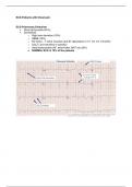
ECG Patients with Chest pain
ECG Pulmonary Embolism
Sinus tachycardia (44%)
Sometimes
o Right axis deviation (16%)
o RBBB (18%)
o RV strain – T wave inversion and ST depression in V1, V2, V3, V4 (34%)
o SIQIIITIII (not sensitive or specific)
o Atrial tachycardias (AF, atrial flutter, MAT etc) (8%)
o NORMAL ECG in 18% of the patients
, Pericarditis ECG features
Wide spread ST elevation concave upwards
Wide spread PR depression = Most specific finding
Reversed in right sided leads aVR and V1 (reciprocal changes)
Changes are usually small (0.5-1mm)
Above Acute changes might be followed by
T wave flattening
T wave inversion
Normalisation of the ECG
Left ventricular hypertrophy (LVH)
S wave depth in V1+Tallest R wave in V5 and V6 >35mm (7 big squares)
Left axis deviation
LVH non-voltage criteria
LV strain ST depression and inverted T waves in left leads
Increased R wave peak time in V5 and V6 (50ms)