Summary Human anatomy & physiology
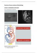
Lecture 1: Introduction to the heart
From a cardiomyocyte to a heart beat
A tale of two circulations
Pulmonary circulation > low pressure system
Systemic circulation> high pressure
➢ Thickness of the wall
,Function of the heart
• Pumping deoxygenated blood to the lungs
• Pumping oxygenated blood to all the organs in the body
• Together with blood vessels: providing adequate perfusion of all organs & tissues of the
body
• Contraction and relaxation determine cardiac output (and the amount of blood)
• How can they be sustained →
– Coordination of contraction and relaxation of 2-3 billion CMs
Excitation-contraction coupling (=action potential at the cellular level)
Contraction of the heart following electrical stimulation of cardiomyocytes
Automation of the heart
• The heart can beat independent of hormonal or neuronal input (but there can be input)
AUTOMATION
• Spontaneous active
• Pacemaker cells (SA node> heart rhythm)
Conduction through the heart
You don’t want your
atria and ventricles
to contract
simultaneously, thus
slow conduction AV
node.
So, first atria
contracts, and then
after being filled with
blood, the ventricles
,Nerve cells in the heart give fast signals to the ventricles
Conduction between cardiomyocytes (electric coupled, slow conduction)
, Action potentials in cardiomyocytes
Can you deduce why automation of heart beat occurs from SA node cells?
• Unstable resting potential • Stable resting potential: -85 mV
• Slow depolarisation prepotential • Quick depolarisation
(pacemaker potential) • Plateau
• Quick repolarisation
Basis for the resting membrane potential
Membrane potential
determined by:
- concentrations differences of ions
AND permeability to ions
-Largely determined by K+ gradient
(see Nernst equation)> high inside,
low outside
Cell in rest is permeable to K+
Cell is slightly negatively charged
Ion channels open and close & action potential of ventricular cell