EPIDERMAL STEM CELLS
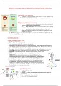
Human Skin
* Largest organ in the body - 12-15% body weight, 1.6 trillion cells • Interface with the external
environment
* Dynamic tissue with important physiological functions
* Undergoes constant turnover - Dust is actually 75% to 90% dead skin cells
* Affected by common and rare diseases - Most common reason for visiting a GP
Why Use Skin as a Model for Stem Cell Biology & Tissue
Morphogenesis?
Readily accessible and abundant.
All derived from a common embryonic skin progenitor cell.
Adult skin epithelia undergo constant turnover in homeostasis and wound
repair.
Epithelial stem cells of the skin can be cultured without losing stemness.
Skin cancers are the most common cancers.
Goal: to understand mechanisms that regulate
adult epithelial stem cells – under homeostatic,
wound-healing and pathological conditions.
o Where are the stem cells located?
o What factors regulate self-renewal and exit from the
stem cell compartment?
o How is choice of lineage controlled?
o How do the stem cells contribute to wound healing
and tumour development?
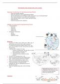
Emergence of distinct stem cell populations
during morphogenesis
Initially, various stem cell markers are coexpressed
within the same region
At later stages marker expression is associated with
segregation of cells into distinct domains.
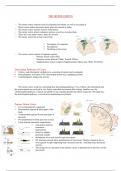
Activation of Hair Growth
- Quiescent stem cells (green) receive inhibitory signals from their differentiated
progeny (red).
- During the resting phase, crosstalk between the mesenchymal cells (purple) and
“primed” stem cells (blue) builds up the threshold of activating signals that
overpower the inhibitory signals to launch the tissue-generating phase.
- The primed stem cells begin to make short-lived progeny (orange), which act as
a transient signaling center to fuel tissue growth.
, Label-retaining cells reside in the bulge area of pilosebaceous unit
* Challenged view that hair follicle stem cells reside in the matrix area of the hair
bulb.
* The bulge consists of a subpopulation of outer root sheath cells located in the
midportion of the follicle at the arrector pili muscle attachment site.
* Normally slow cycling,but can be stimulated to proliferate transiently.
* This new view provides insights into hair cycle control and the possible
involvement of hair follicle stem cells in skin carcinogenesis.
Involvement of Follicular Stem Cells in Forming Not Only the
Follicle but Also the Epidermis
Hair follicular stem cells, located in the bulge region, can give rise
to several cell types of the hair follicle as well as upper follicular
cells.
Upper follicular keratinocytes migrate into the epidermis in normal newborn mouse
skin, and in adult mouse skin in response to a penetrating wound.
Follicular bulge stem cells are potentially bipotent as they can give rise to not only the
hair follicle, but also the epidermis.
CD34 is a specific marker of bulge cell keratinocytes
o Facilitates isolation of live epithelial cells with
stem/progenitor cell characteristics
o Potentially providing a tool for the study of carcinogen target
cells, gene therapy, and tissue engineering applications.
Hair Follicle Stem Cell Compartments
o Each SC compartment is responsible for sustaining tissue homeostasis within a
discrete skin domain. (details in table later in talk)
o When challenged via wounding or removed from their native niche and
engrafted, epithelial SCs are often less restricted in their fates. Cell plasticity
Human Skin
* Largest organ in the body - 12-15% body weight, 1.6 trillion cells • Interface with the external
environment
* Dynamic tissue with important physiological functions
* Undergoes constant turnover - Dust is actually 75% to 90% dead skin cells
* Affected by common and rare diseases - Most common reason for visiting a GP
Why Use Skin as a Model for Stem Cell Biology & Tissue
Morphogenesis?
Readily accessible and abundant.
All derived from a common embryonic skin progenitor cell.
Adult skin epithelia undergo constant turnover in homeostasis and wound
repair.
Epithelial stem cells of the skin can be cultured without losing stemness.
Skin cancers are the most common cancers.
Goal: to understand mechanisms that regulate
adult epithelial stem cells – under homeostatic,
wound-healing and pathological conditions.
o Where are the stem cells located?
o What factors regulate self-renewal and exit from the
stem cell compartment?
o How is choice of lineage controlled?
o How do the stem cells contribute to wound healing
and tumour development?
Emergence of distinct stem cell populations
during morphogenesis
Initially, various stem cell markers are coexpressed
within the same region
At later stages marker expression is associated with
segregation of cells into distinct domains.
Activation of Hair Growth
- Quiescent stem cells (green) receive inhibitory signals from their differentiated
progeny (red).
- During the resting phase, crosstalk between the mesenchymal cells (purple) and
“primed” stem cells (blue) builds up the threshold of activating signals that
overpower the inhibitory signals to launch the tissue-generating phase.
- The primed stem cells begin to make short-lived progeny (orange), which act as
a transient signaling center to fuel tissue growth.
, Label-retaining cells reside in the bulge area of pilosebaceous unit
* Challenged view that hair follicle stem cells reside in the matrix area of the hair
bulb.
* The bulge consists of a subpopulation of outer root sheath cells located in the
midportion of the follicle at the arrector pili muscle attachment site.
* Normally slow cycling,but can be stimulated to proliferate transiently.
* This new view provides insights into hair cycle control and the possible
involvement of hair follicle stem cells in skin carcinogenesis.
Involvement of Follicular Stem Cells in Forming Not Only the
Follicle but Also the Epidermis
Hair follicular stem cells, located in the bulge region, can give rise
to several cell types of the hair follicle as well as upper follicular
cells.
Upper follicular keratinocytes migrate into the epidermis in normal newborn mouse
skin, and in adult mouse skin in response to a penetrating wound.
Follicular bulge stem cells are potentially bipotent as they can give rise to not only the
hair follicle, but also the epidermis.
CD34 is a specific marker of bulge cell keratinocytes
o Facilitates isolation of live epithelial cells with
stem/progenitor cell characteristics
o Potentially providing a tool for the study of carcinogen target
cells, gene therapy, and tissue engineering applications.
Hair Follicle Stem Cell Compartments
o Each SC compartment is responsible for sustaining tissue homeostasis within a
discrete skin domain. (details in table later in talk)
o When challenged via wounding or removed from their native niche and
engrafted, epithelial SCs are often less restricted in their fates. Cell plasticity