AORTIC DISSECTION
Aortic dissection = disruption of medial layer of aorta (tunica media) due to blood, causing separation of the layers,
resulting in a true lumen and false lumen. It is a medical emergency.
Acute aortic syndrome = disruption of the arterial wall layers, split into 3 subgroups: aortic dissection, penetrating aortic
ulcer, intramural hematoma.
-----------------------------------------------------------------------------------------------------------------------------------------------------
Presentation
§ Chest pain ® tearing in nature
§ Abdominal pain
§ Pain radiating to the back
§ Dyspnea
§ Syncope
§ Tachycardia
§ Hypotension
§ New aortic regurgitation murmur
§ Intra-arm blood pressure differential
§ Radial pulse deficit
§ Neurological deficit
§ Horner’s syndrome
§ Absent peripheral pulses
§ Signs of end-organ hypoperfusion ® reduced urine output, paraplegia, lower limb ischaemia, abdominal pain 2nd to
ischaemia, deteriorating conscious level
-----------------------------------------------------------------------------------------------------------------------------------------------------
Pathophysiology
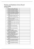
Disruption of aortic wall due to blood, causing separation of the layers, resulting in a true lumen and false lumen.
§ Artery walls consist of three main layers: tunica intima (innermost) ® tunica media ® tunica adventitia (outermost)
§ Aortic dissection is a tear in the intimal layer of the aortic wall
o Blood flows between this layer and splits apart the tunica intima and media
o This creates a true lumen and a false lumen
§ The tear can progress distally, proximally or in both directions from origin
o Anterograde dissection ® towards iliac arteries
o Retrograde dissection ® towards aortic valve
§ Can result in prolapse of aortic valve, bleeding into pericardium and cardiac tamponade
§ Acute = diagnosed <14 days after occurrence
§ Chronic = diagnosed >14 days after occurance
§ Other conditions that fall under the acute aortic syndrome can cause a dissection eventually, including:
o Penetrating aortic ulcer: penetrates the intima and progresses into the media of artery
o Intramural hematoma: contained aortic wall hematoma with bleeding into the media.
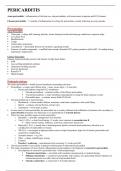
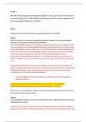
Stanford Classification
Divides aortic dissection into two groups, A and B:
§ Type A ® involves the ascending aorta and can propagate to the aortic arch and descending aorta (DeBakey Types I
and II), the tear can originate anywhere along this path
§ Type B ® does not involve the ascending aorta, occurring in any other part of the aortic arch and descending aorta
(DeBakey Type III).
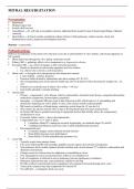
DeBakey Classification
This groups dissections anatomically.
§ Type I ® originates in ascending aorta and propagates to at least the aortic arch
o Typically seen in patients <65 and carry highest mortality.
Aortic dissection = disruption of medial layer of aorta (tunica media) due to blood, causing separation of the layers,
resulting in a true lumen and false lumen. It is a medical emergency.
Acute aortic syndrome = disruption of the arterial wall layers, split into 3 subgroups: aortic dissection, penetrating aortic
ulcer, intramural hematoma.
-----------------------------------------------------------------------------------------------------------------------------------------------------
Presentation
§ Chest pain ® tearing in nature
§ Abdominal pain
§ Pain radiating to the back
§ Dyspnea
§ Syncope
§ Tachycardia
§ Hypotension
§ New aortic regurgitation murmur
§ Intra-arm blood pressure differential
§ Radial pulse deficit
§ Neurological deficit
§ Horner’s syndrome
§ Absent peripheral pulses
§ Signs of end-organ hypoperfusion ® reduced urine output, paraplegia, lower limb ischaemia, abdominal pain 2nd to
ischaemia, deteriorating conscious level
-----------------------------------------------------------------------------------------------------------------------------------------------------
Pathophysiology
Disruption of aortic wall due to blood, causing separation of the layers, resulting in a true lumen and false lumen.
§ Artery walls consist of three main layers: tunica intima (innermost) ® tunica media ® tunica adventitia (outermost)
§ Aortic dissection is a tear in the intimal layer of the aortic wall
o Blood flows between this layer and splits apart the tunica intima and media
o This creates a true lumen and a false lumen
§ The tear can progress distally, proximally or in both directions from origin
o Anterograde dissection ® towards iliac arteries
o Retrograde dissection ® towards aortic valve
§ Can result in prolapse of aortic valve, bleeding into pericardium and cardiac tamponade
§ Acute = diagnosed <14 days after occurrence
§ Chronic = diagnosed >14 days after occurance
§ Other conditions that fall under the acute aortic syndrome can cause a dissection eventually, including:
o Penetrating aortic ulcer: penetrates the intima and progresses into the media of artery
o Intramural hematoma: contained aortic wall hematoma with bleeding into the media.
Stanford Classification
Divides aortic dissection into two groups, A and B:
§ Type A ® involves the ascending aorta and can propagate to the aortic arch and descending aorta (DeBakey Types I
and II), the tear can originate anywhere along this path
§ Type B ® does not involve the ascending aorta, occurring in any other part of the aortic arch and descending aorta
(DeBakey Type III).
DeBakey Classification
This groups dissections anatomically.
§ Type I ® originates in ascending aorta and propagates to at least the aortic arch
o Typically seen in patients <65 and carry highest mortality.