Secondary structure of Nucleic acid
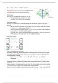
Structure of the duplex DNA
• Right-handed double helix
• 2 nucleotide chain running anti-parallel to one another
• 5' to 3' directionality
• 2 chains interact by the bases forming complementary base pairing (A-T and G-C)
• 1 helical turn (1 pitch) = 34 Å (3.4 nm)
• Width = 2 nm
• Distance btwn 2 bases (vertical) = 3.4 Å (0.34 nm)
Base stacking
• Occurs perpendicular to the helical axis
• Flat bases stack on top of each other
• Electron cloud (pi-pi) interaction from the aromatic ring between the stacked bases contribute to the stability
• Stacked bases attracted by induced dipole (van der Waalsforce) between the electron clouds
• Base stacking also contributes to hydrophobic effect
• Hydrophobic bases are buried by base stacking
Major and Minor groove
·
• Why do grooves exist? - the geometry of the base pair / the angle of the
glycosidic bonds is 120° (narrow angle) or 240° (wide angle)
• Minor groove: narrow angles on one edge of the base pairs
• Major groove: wide angles on the other edge
The major groove is rich in information
• H-bond acceptor (A), H-bond donator (D), Methyl group (M), Non-polar
hydrogen (H)
• Edge of A=T: ADAM signify AT base pair in the major groove
• Edge of G≡C: AADH stands for GC base pair
• It allow proteins to bind to specific DNA sequences without directly
interacting with the bases and disrupting the structure
Watson-Crick base pairing
• Derives from: complementarity of shape and hydrogen bonding properties of the 4
bases
• A=T H-bond between: C6 amino group (exocyclic) A + C4 carbonyl group T / N1 of A +
N3 of T
• G≡C H-bond between: C2 NH2 (exocyclic) on G + C2 carbonyl on C / N1 on G + N3 on
C / C6 carbonyl on G + C4 NH2 (exocyclic) on C
Geometry
• 2 base pairs have exactly the same geometry → distances between 2 sugars are the
same = symmetry
• All 4 bases are accommodated without distorting the DNA structure
• Base pairs can stack on top of each other
• Order of bases are irregular but the overall DNA structure is regular
Chargaff’s Rule
• The relative ratios of the 4 bases are not random
• The number of bases A = T, G=C
• Regardless of the DNA source, purine: pyrimidine = 1:1 (ratio of 1)
• Keto bases : Amino bases = 1 : 1
Requirements for Watson-Crick base pairing
• Each of the 4 bases exist in 2 alternative tautomeric states in an equilibrium
• Nitrogen on amino base: predominantly NH2 forms and rarely the imino (H-N=C) forms
• Oxygen on keto bases: predominantly C=O forms and rarely the enol (C-O-H) forms
• Frequent source of errors during DNA synthesis
Different forms of DNA
1. B-form
• Right handed
• Rises between adjacent bases = 0.34nm
• Helical repeat = 3.4nm
• 10 bp/ turn, 2 nm wide
• Distances vary because DNA is dynamic and a moving molecule → B form is an AVERAGE structure
• DNA is more stretched out in solution = 10.4 bp/ turn