Topic 1: Cells
Get ready to explore the world of…
Microscopes Animal cell
Animal and plant cells
Prokaryotic and Eukaryotic cells
Specialisation in animal cells
Specialisation in plant cells
Diffusion
Osmosis plant cell
Active transport
Exchanging materials
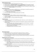
Summary
● There are two types of cells: prokaryotic cells and eukaryotic cells. Prokaryotic cells are cells that do not have a nucleus,
whereas eukaryotic cells have one. An example of a prokaryotic cell is bacteria and examples of eukaryotic cells include animal
and plant cells.
● Animal and Plant cells both have a nucleus, cell membrane, cytoplasm and mitochondria. Animal cells have ribosomes, which
plant cells don’t have and plant cells have a cell wall, chloroplasts and a permanent vacuole that animal cells do not have.
● Bacteria have a cell wall, a cell membrane, chromosomal DNA, plasmid DNA, cytoplasm and a flagellum.
● There are two types of microscopes: the light microscope and the electron microscope. The electron microscope has both
higher magnification and resolution than the light microscope; If you are asked to calculate the magnification of an image,
you use the formula Magnification = image size/real size.
● Not all animal and plant cells are the same. That is because cells are specialised for their specific function. The process of a
cell becoming specialised is called differentiation. A cell that has not yet been specialised is called a stem cell.
● Cells in multi-cellular organisms go through the cell cycle, which involves growth and DNA replication & finally mitosis.
Mitosis is the process in which cells duplicate and reproduce.
● Diffusion is the movement of particles from an area of high concentration to an area of low concentration. Osmosis is a
type of diffusion that happens in water - osmosis is the movement of particles from a high water concentration to a low
water concentration across a partially permeable membrane. Active transport is the movement of particles against a
concentration gradient - from a low concentration to a high concentration.
● Most exchange surfaces are adapted with 3 characteristics: they have a thin membrane, large surface area and a good blood
supply.
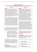
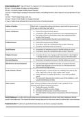
, Microscopes
There are two types of microscopes: the Light Microscope and the Electron Microscope.
The light microscope was invented in the mid-17th century and it uses a beam of light or a
mirror (where the light would be reflected) to show us the cells. These are the type of
microscopes you’d find at school. They have a magnification of x2000 and a resolution of
200 nm.
Electron microscopes were invented in the 1930s and they use a beam of electrons to show
us the sub-cellular parts of the cells. These helped scientists learn more about parts as
small as ribosomes, that you could hardly see in light microscopes. Electron microscopes are
more expensive than light microscopes and they have a magnification of x2 000 000 and a
resolution of 0.2 nm.
Magnification is how big the image is and the resolution is how clear the image is.
In the exam, you may be asked to calculate the magnification of an image. To do so, you
can use the formula Magnification = Image size/Real size.
If you are asked to find the total magnification, you will just simply multiply the
magnification of the eyepiece lens by the magnification of the objective lens.
Questions
1) Outline 3 advantages of electron microscopes. (3)
2) Compare light microscopes to electron microscopes. (4)
3) A student looked at a cell with a diameter of 10000 nanometres. He then measured the diameter
of the cell under the microscope and it was 5000 nanometers. Calculate the magnification. (2)
Get ready to explore the world of…
Microscopes Animal cell
Animal and plant cells
Prokaryotic and Eukaryotic cells
Specialisation in animal cells
Specialisation in plant cells
Diffusion
Osmosis plant cell
Active transport
Exchanging materials
Summary
● There are two types of cells: prokaryotic cells and eukaryotic cells. Prokaryotic cells are cells that do not have a nucleus,
whereas eukaryotic cells have one. An example of a prokaryotic cell is bacteria and examples of eukaryotic cells include animal
and plant cells.
● Animal and Plant cells both have a nucleus, cell membrane, cytoplasm and mitochondria. Animal cells have ribosomes, which
plant cells don’t have and plant cells have a cell wall, chloroplasts and a permanent vacuole that animal cells do not have.
● Bacteria have a cell wall, a cell membrane, chromosomal DNA, plasmid DNA, cytoplasm and a flagellum.
● There are two types of microscopes: the light microscope and the electron microscope. The electron microscope has both
higher magnification and resolution than the light microscope; If you are asked to calculate the magnification of an image,
you use the formula Magnification = image size/real size.
● Not all animal and plant cells are the same. That is because cells are specialised for their specific function. The process of a
cell becoming specialised is called differentiation. A cell that has not yet been specialised is called a stem cell.
● Cells in multi-cellular organisms go through the cell cycle, which involves growth and DNA replication & finally mitosis.
Mitosis is the process in which cells duplicate and reproduce.
● Diffusion is the movement of particles from an area of high concentration to an area of low concentration. Osmosis is a
type of diffusion that happens in water - osmosis is the movement of particles from a high water concentration to a low
water concentration across a partially permeable membrane. Active transport is the movement of particles against a
concentration gradient - from a low concentration to a high concentration.
● Most exchange surfaces are adapted with 3 characteristics: they have a thin membrane, large surface area and a good blood
supply.
, Microscopes
There are two types of microscopes: the Light Microscope and the Electron Microscope.
The light microscope was invented in the mid-17th century and it uses a beam of light or a
mirror (where the light would be reflected) to show us the cells. These are the type of
microscopes you’d find at school. They have a magnification of x2000 and a resolution of
200 nm.
Electron microscopes were invented in the 1930s and they use a beam of electrons to show
us the sub-cellular parts of the cells. These helped scientists learn more about parts as
small as ribosomes, that you could hardly see in light microscopes. Electron microscopes are
more expensive than light microscopes and they have a magnification of x2 000 000 and a
resolution of 0.2 nm.
Magnification is how big the image is and the resolution is how clear the image is.
In the exam, you may be asked to calculate the magnification of an image. To do so, you
can use the formula Magnification = Image size/Real size.
If you are asked to find the total magnification, you will just simply multiply the
magnification of the eyepiece lens by the magnification of the objective lens.
Questions
1) Outline 3 advantages of electron microscopes. (3)
2) Compare light microscopes to electron microscopes. (4)
3) A student looked at a cell with a diameter of 10000 nanometres. He then measured the diameter
of the cell under the microscope and it was 5000 nanometers. Calculate the magnification. (2)