TOKLA – NEUROANATOMIE
FUNCTIONEEL
Functionele anatomie: somatisch afferent
Functioneel is het in 2 groepen te verdelen: afferent en efferent
• Afferent = info aangevoerd naar centraal vanuit perifeer
• Efferent = info die van perifeer naar centraal gaat
Somatisch afferent
= info die komt vanuit lichaam zelf – meestal bewust
2 grote systemen: lemniscaal en extralemniscaal systeem
Lemniscaal systeem
• Vnl belang bij primaten en mens – huisdieren minder sterk
• Fijn tastgevoel, transporteren prikkels, druk, vibraties (specifiek)
• Bewuste proprioceptie
• Zeer snel systeem
• Geen spinale interneuronen, geen synaps
• Zintuiglijke waarnemingen en proprioceptie getransporteerd
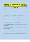
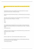
Info komt vanuit perifeer à naar RM à geen contact met dorsale hoorn,
maar direct naar dorsale funniculus à ascenderen
Primaire neuronen
• Fasciculus gracilis: loopt dichtst med en komt vanuit achterhand
• Fasciculus cuneatus: veel groter en komt vanuit voorhand
Secundaire neuronen
• Nucleus gracilis
• Nucleus cuneatus
à vormen de mediale lemniscus (contralateraal)
-loopt ventraal, steekt over en eindigt thv thalamus (caud)
Tertiaire neuronen
• Thalamus: caudoventrale nucleus
• Via capsula interna naar somatosensorsich gebied cortex (neo)
Proces
1° gaan ascenderen à MO à nucleus gracilis en cuneatus à synaps: 2° à
mediale lemniscus gevormd à thalamus à 3° neuronen à sensibele/
sensorische cortex
Extralemniscaal systeem
• Vnl bij onze huisdieren
• Transporteren van ruw tastgevoel, durk, T, vibraties, pijn
• Informatie is minder precies
• Wel spinale interneuronen, wel synaps thv RM
, Secundaire neuronen
In RM vertrekken 2° neuronen à ascenderen – kan op versch manieren
• Bundelen in laterale groep
• Bundelen in mediale groep
1) Bundelen in laterale groep
a) zwarte lijnen: steken over à naar controlateraal à vormt meer ventr in
laterale funniculus een tractus spinothalamicus (of neo) à ascenderen à
contact met thalamus thv med caudoventrale kernen in thalamus
• Tractus neospinothalamicus transporteert vnl pijnvezels
b) witte lijn: blijft ipsilateraal à vormt tractus spinocervicothalamicus, iets
meer dors in laterale funniculus à maakt synaps thv MO à steekt over à
contralateraal à contact met med caudoventrale kernen in thalamus à
via capsula interna naar sensorische cortex
c) second order dorsal column pathway: specifiek bij kat
2) Bundelen in mediale groep
• Aantal synapsen op verloop van mediale groep
• Alleen in SG
a) info komt binnen à vanuit dors hoorn à blijven in dors hoorn à naar
rostraal à maken versch keren achter elkaar synapsen met rostr
segmenten à grijze hoorn verlaten à in SA à vormen tractus
paleospinothalamicus (ipsi en contralat) à eindigen in thalamische nuclei
(mediale en interlaminaire) à capsula interna à sensorische cortex
b) ractus spinoreticularis (bilat) verlaat grijze hoorn à SA à naar formatio
reticularis
c) diffusie ascenderende vezels: lopen in SA à vormen niet echt tractus
• Iets te maken met proprioceptie (bewust propriospinaal systeem)
Lemniscaal en extralemniscaal systeem: samengevat
Bewust en finaal contralateraal verlopend naar sensorische cortex
On(der)bewuste proprioceptie
• Vanuit lichaam zelf, spieren en pezen: onbewust
Binnen via spinale ganglia vanuit pees- en spierspoeltjes à contact maken
in dorsale hoornen à 2° neuronen à bundelen in SA à spinocerebellaris
dorsalis en tractus spinocerebellaris ventralis in lat funniculus perifeer
1) Tractus spinocerebellaris dorsalis
• Info vanuit spierspoeltjes
• Blijft ipsilteraal verlopen – kruist niet thv RM
• 2° neuronen maken geen synaps meer
• Beweging heel vlot laten gebeuren
Treden binnen via caud cerebellaire pedunkel naar cerebellaire cortex à
info aangevoerd in cerebellum à al dan niet stimuleren purkinjecellen
FUNCTIONEEL
Functionele anatomie: somatisch afferent
Functioneel is het in 2 groepen te verdelen: afferent en efferent
• Afferent = info aangevoerd naar centraal vanuit perifeer
• Efferent = info die van perifeer naar centraal gaat
Somatisch afferent
= info die komt vanuit lichaam zelf – meestal bewust
2 grote systemen: lemniscaal en extralemniscaal systeem
Lemniscaal systeem
• Vnl belang bij primaten en mens – huisdieren minder sterk
• Fijn tastgevoel, transporteren prikkels, druk, vibraties (specifiek)
• Bewuste proprioceptie
• Zeer snel systeem
• Geen spinale interneuronen, geen synaps
• Zintuiglijke waarnemingen en proprioceptie getransporteerd
Info komt vanuit perifeer à naar RM à geen contact met dorsale hoorn,
maar direct naar dorsale funniculus à ascenderen
Primaire neuronen
• Fasciculus gracilis: loopt dichtst med en komt vanuit achterhand
• Fasciculus cuneatus: veel groter en komt vanuit voorhand
Secundaire neuronen
• Nucleus gracilis
• Nucleus cuneatus
à vormen de mediale lemniscus (contralateraal)
-loopt ventraal, steekt over en eindigt thv thalamus (caud)
Tertiaire neuronen
• Thalamus: caudoventrale nucleus
• Via capsula interna naar somatosensorsich gebied cortex (neo)
Proces
1° gaan ascenderen à MO à nucleus gracilis en cuneatus à synaps: 2° à
mediale lemniscus gevormd à thalamus à 3° neuronen à sensibele/
sensorische cortex
Extralemniscaal systeem
• Vnl bij onze huisdieren
• Transporteren van ruw tastgevoel, durk, T, vibraties, pijn
• Informatie is minder precies
• Wel spinale interneuronen, wel synaps thv RM
, Secundaire neuronen
In RM vertrekken 2° neuronen à ascenderen – kan op versch manieren
• Bundelen in laterale groep
• Bundelen in mediale groep
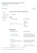
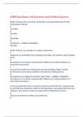
1) Bundelen in laterale groep
a) zwarte lijnen: steken over à naar controlateraal à vormt meer ventr in
laterale funniculus een tractus spinothalamicus (of neo) à ascenderen à
contact met thalamus thv med caudoventrale kernen in thalamus
• Tractus neospinothalamicus transporteert vnl pijnvezels
b) witte lijn: blijft ipsilateraal à vormt tractus spinocervicothalamicus, iets
meer dors in laterale funniculus à maakt synaps thv MO à steekt over à
contralateraal à contact met med caudoventrale kernen in thalamus à
via capsula interna naar sensorische cortex
c) second order dorsal column pathway: specifiek bij kat
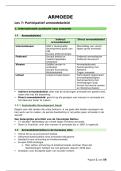
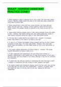
2) Bundelen in mediale groep
• Aantal synapsen op verloop van mediale groep
• Alleen in SG
a) info komt binnen à vanuit dors hoorn à blijven in dors hoorn à naar
rostraal à maken versch keren achter elkaar synapsen met rostr
segmenten à grijze hoorn verlaten à in SA à vormen tractus
paleospinothalamicus (ipsi en contralat) à eindigen in thalamische nuclei
(mediale en interlaminaire) à capsula interna à sensorische cortex
b) ractus spinoreticularis (bilat) verlaat grijze hoorn à SA à naar formatio
reticularis
c) diffusie ascenderende vezels: lopen in SA à vormen niet echt tractus
• Iets te maken met proprioceptie (bewust propriospinaal systeem)
Lemniscaal en extralemniscaal systeem: samengevat
Bewust en finaal contralateraal verlopend naar sensorische cortex
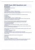
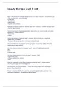
On(der)bewuste proprioceptie
• Vanuit lichaam zelf, spieren en pezen: onbewust
Binnen via spinale ganglia vanuit pees- en spierspoeltjes à contact maken
in dorsale hoornen à 2° neuronen à bundelen in SA à spinocerebellaris
dorsalis en tractus spinocerebellaris ventralis in lat funniculus perifeer
1) Tractus spinocerebellaris dorsalis
• Info vanuit spierspoeltjes
• Blijft ipsilteraal verlopen – kruist niet thv RM
• 2° neuronen maken geen synaps meer
• Beweging heel vlot laten gebeuren
Treden binnen via caud cerebellaire pedunkel naar cerebellaire cortex à
info aangevoerd in cerebellum à al dan niet stimuleren purkinjecellen