Lecture 8
Bone development, Growth and Repair
- Bones forms by replacing existing connective tissues in the fetus
o Intramembranous bones
Develop from sheet-like layers of undifferentiated connective tissue
Turn into a cell type -> Osteoblasts -> bone builder
Osteoclasts -> bone breaker
Osteoblasts deposit a bony matrix around themselves in all directions
forming spongy bone
Osteocytes is the mature type of Osteoblasts
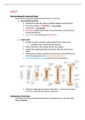
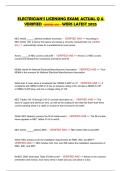
o Endochondral
Develop as hyaline cartilage models shaped like the future bones
Cartilage is then replaced with bone tissues
Most of the bone of the skeleton fall into this category
Growth plate (Epiphyseal plates) is the place allow the bone to grow
longer
Disintegrating cartilage is invaded by Blood vessels and osteoblasts that
first form spongy bone at the primary ossification center
Secondary ossification center appears later in the epiphyses
On X-ray: cartage will not show in white colour -> so if do not see gap in
x-ray, we can tell that this could be a grown-up
Homeostasis of Bone Tissue
- Osteoclasts resorb and osteoblasts deposit bone throughout life, in a process called
bone remodeling
, - An average of 3% to 5% of bone calcium is exchanged each year
- The remodeling process is controlled by hormones that regulate blood calcium level
Factoring affecting Bone development
- Nutrition: Vitamin D is needed for Ca absorption
o By Food
o By supplement
- Hormonal secretions:
o Growth hormone – stimulates division of cartilage cells of the epiphyseal plate
o Sex hormone – stimulate ossification of the epiphyseal plates to end growth in
height
- Physical exercise: muscle pull on bones at attachment sites, stress the bone -> increase
thickness and strength
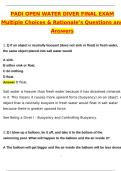
Skeletal organization
- Axial skeleton – consists of the bony and cartilaginous parts that support the and
protect the head, neck and trunk
o Skull: cranium and facial bones
o Hyoid bone: supports the tongue and aids in swallowing
o Vertebral column
o Thoracic cage: ribs and sternum
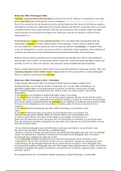
- Appendicular skeleton – consists of the bones of the upper and
lower limbs
o Pectoral girdle: clavicle and scapula
o Upper limbs: humerus, ulna, radius, carpals, metacarpals,
and phalanges
o Pelvic girdle: 2 hip bones
o Lower limbs: femur, patella, tibia, fibula, tarsals, metatarsals, and phalanges
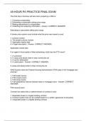
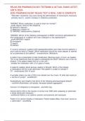
Skull
- Made up of 22 bones
- Provide attachments for muscles and contains air filled paranasal sinuses that reduce its
weight and increase vocal resonance
o 8 cranial bones *Cranium bone – encloses and protects the brain*
Frontal bone (include the supraorbital foramen)
(coronal suture)
Parietal bones x2 (Squamous suture) Temporal bone x2
(lambdoid suture)
Occipital bone
Bone development, Growth and Repair
- Bones forms by replacing existing connective tissues in the fetus
o Intramembranous bones
Develop from sheet-like layers of undifferentiated connective tissue
Turn into a cell type -> Osteoblasts -> bone builder
Osteoclasts -> bone breaker
Osteoblasts deposit a bony matrix around themselves in all directions
forming spongy bone
Osteocytes is the mature type of Osteoblasts
o Endochondral
Develop as hyaline cartilage models shaped like the future bones
Cartilage is then replaced with bone tissues
Most of the bone of the skeleton fall into this category
Growth plate (Epiphyseal plates) is the place allow the bone to grow
longer
Disintegrating cartilage is invaded by Blood vessels and osteoblasts that
first form spongy bone at the primary ossification center
Secondary ossification center appears later in the epiphyses
On X-ray: cartage will not show in white colour -> so if do not see gap in
x-ray, we can tell that this could be a grown-up
Homeostasis of Bone Tissue
- Osteoclasts resorb and osteoblasts deposit bone throughout life, in a process called
bone remodeling
, - An average of 3% to 5% of bone calcium is exchanged each year
- The remodeling process is controlled by hormones that regulate blood calcium level
Factoring affecting Bone development
- Nutrition: Vitamin D is needed for Ca absorption
o By Food
o By supplement
- Hormonal secretions:
o Growth hormone – stimulates division of cartilage cells of the epiphyseal plate
o Sex hormone – stimulate ossification of the epiphyseal plates to end growth in
height
- Physical exercise: muscle pull on bones at attachment sites, stress the bone -> increase
thickness and strength
Skeletal organization
- Axial skeleton – consists of the bony and cartilaginous parts that support the and
protect the head, neck and trunk
o Skull: cranium and facial bones
o Hyoid bone: supports the tongue and aids in swallowing
o Vertebral column
o Thoracic cage: ribs and sternum
- Appendicular skeleton – consists of the bones of the upper and
lower limbs
o Pectoral girdle: clavicle and scapula
o Upper limbs: humerus, ulna, radius, carpals, metacarpals,
and phalanges
o Pelvic girdle: 2 hip bones
o Lower limbs: femur, patella, tibia, fibula, tarsals, metatarsals, and phalanges
Skull
- Made up of 22 bones
- Provide attachments for muscles and contains air filled paranasal sinuses that reduce its
weight and increase vocal resonance
o 8 cranial bones *Cranium bone – encloses and protects the brain*
Frontal bone (include the supraorbital foramen)
(coronal suture)
Parietal bones x2 (Squamous suture) Temporal bone x2
(lambdoid suture)
Occipital bone