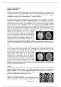
Lecture 1 part 1
Examples of viruses
➢ Smallpox
○ very old virus
○ Eradicated by vaccination (1967-1980 campaign)
➢ Poliomyelitis
○ very old virus
○ Caused by poliovirus, belonging to the family Picornaviridae
○ course of infection
■ infection via mouth
■ replication in the small intestine
■ Lymphnodes and bloodstream
■ Nerve infection: replication, intraneuronal spread
■ Excretion of virus in feces
○ paralysis, death, iron lungs
➢ Rinderpest
○ 90% of cattle died
➢ Measles
➢ Herpes virus infections
○ Chickenpox
○ shingles (girdle herpes)
○ fever blisters
➢ Human papillomavirus
○ HPV (genital) wart virus
○ Sexually transmitted
○ Some variants however cause (cervical) cancer
➢ HIV
○ It can lead to AIDS
○ Acquired Immuno Deficiency Syndrome: The virus targets the immune
system
General information viruses
➢ Virus = Smallest genetic entitiy
➢ All organisms have viruses
➢ Most viruses are found in surface water
➢ Adolf Mayer → found the first virus → tobacco mosaic disease
➢ very big variance in the genome sizes
inert
= inactive / non-moving
,The minimal virus
● DNA‐ or RNA‐polymerase → multiplication of RNA or DNA genome
● Coat or capsid protein → protection or host interactions
Shapes
Viral envelope
= A lipid bilayer/membrane with transmembrane proteins (spikes) pointing
outwards
, Lecture 1 part 2
Methods used in virology
Methods for generation and quantification of viruses
➢ Cultivation in host
○ whole organism
○ embryonated eggs
○ cell culture
→ Types of cell culture: primary culture and
immortalize line
➢ Isolation
○ Plaque purification (also used for quantification)
○ Centrifugation → Differential (low/high speed) or
density gradient (sucrose)
➢ Quantification
○ Real-time PCR
○ EPDA (End Point Dilution assay): TCID50 assay
Methods for structural investigation
➢ Localization and accumulation
○ Light microscopy → stain
○ Fluorescence microscopy → GFP fused protein
➢ Structure (3D)
○ Electron microscopy (scanning and transmission)
○ X-ray crystallography
○ NMR (nuclear magnetic resonance)
○ AFM (atomic force microscopy)
Methods for detection
➢ CPE
= cytopathic effect
= structural changes in host cells that are caused by viral invasion
➢ Microscopy-immunodetection
○ Immunostaining, immunofluorescence
○ ELISA
○ Western blot analysis
➢ Antigens-antibodies
○ Hemagglutination assay
= surface projections of viruses leads to
crosslinking of red blood cells. can be quantified
by making serial dilutions of the virus sample
➢ NATs = nucleic acid tests
NA is first extracted and purified, then detected using
PCR