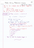
T O P I C A L C A S E 1 – L A C K I N G O X YG E N
learning goals
1. what is hypoxia and how does a hypoxic tumour behave?
2. answer the following questions about the sugar scan:
a) why did they use a sugar scan and what scan did they use?
b) what did they target?
c) what are the results and what do the results of a hypoxia patient look like
when using this scan?
3. answer the following questions about the nitroimidazole scan:
a) why did they use this scan?
b) what did they target?
c) what are the results and what do the results of a hypoxia patient look like
when using this scan?
d) what type of radioactive tracers are used in hypoxic patients?
home study
hypoxia and hypoxic tumours
normally there is a steady state of oxygen concentration in tissues --> balance between
supply and demand. however, sometimes this equilibrium is disturbed and the oxygen
concentration in a tissue becomes too low. the cells of tissue lack oxygen and are in a
state of trying to survive, this is called hypoxia.
o hypoxia = below-normal oxygen level
in normal tissues, blood vessels are arranged in a controlled and systematic manner -->
the distance from the cells to the capillaries establish a constant and uniform oxygen
gradient.
tumour vasculature
abnormal vasculature is the prime cause of hypoxia in tumours
o impaired and chaotic blood vessel development
o tumour cells outgrow the blood supply
o tumour blood vessels are also unstable and might collapse --> causing (temporary)
occlusion
therefore, lack of oxygen supply --> regions of hypoxia [1, 2].
, hypoxia in tumours
Tumour cell hypoxia decreases the effectiveness of anti-cancer treatment and increases
tumour aggressiveness in a number of solid tumours [3]. tumours hypoxia influences
malignancy by adapting via several mechanisms:
o increased anaerobic glycolysis and angiogenesis
o natural selection --> only cells that can cope with low oxygen survive, creating a
more malignant phenotype
o hypoxia can alter the DNA repair capacity --> promoting genomic instability -->
can accelerate cancer development [4]
there are two kinds of hypoxic tumours
1. chronic hypoxia --> diffusion-limited hypoxia
▪ tumour growth cause limited oxygen diffusion
- blood vessels in the tumour are surrounded by proliferating cells
until the diffusion barrier is reached
- the proliferating cells are surrounded by hypoxic cells
2. acute hypoxia --> perfusion-limited hypoxia
▪ temporally unstable blood flow leads to fluctuating perfusion-limited
hypoxia
- (temporary) non-functional blood vessel is surrounded by hypoxic
cells
also, due to the lack of oxygen hypoxic tumours are more resistant to treatment (e.g.
radiotherapy).