Problem 1
Source: Goldstein chapter 2 and 3,
Learning Goals:
• what is the structure of the eye and what are its functions?
• what parts of the eye play a role in perceiving light?
• What conditions/ diseases/ problems are there?
• how do different stimuli trigger our sensory stimuli? how do we perceive different grey
tones?
• How does the variation of light play a role in how we perceive things?
THE VISUAL SYSTEM
Yantis and Abrams Chapter 2
1. Light reflected from objects in the environment and enters the eye through the pupil
2. Focused by the cornea and lens to form sharp images of the objects on the retina which
contains receptors for vision
Shapes and Sizes:
- Spherical à easy to rotate and move
- Optic axis à imaginary diameter line from front to the back
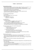
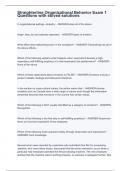
Three membranes:
, - Sclera: protective Outer membrane, made up of (tough protective
covering whose visible portion in the white of eye and transparent
in front of eye)
- Choroid: Middle membrane, lines interior of the sclera, contains
blood vessels that supply inside of eye with oxygen and nutrients
- Retina: inner membrane, made up of neurons, light sensitive
receptors (convert light into neural signals) and other neurons
Cornea: transparent membrane in front of eye
- Light enters the eye by first passing through cornea which
refracts the light
- Focusing process
- Cannot adjust how much light passing through is refracted
Iris: coloured part of eye, muscle with opening, controls size of pupil by contracting and relaxing in
response to intensity of light entering
Pupil: opening through which light enters, size controlled by pupillary light reflex
- Small pupil = it constricts
- Larger = it dilates
Three chambers:
- Anterior chamber: space between the cornea and the iris
o Filled with Aqueous humor
- Posterior chamber: space between lens and iris
o Filled with Aqueous humor
- Vitreous chamber: main interior portion of eye
o Filled with Vitreous humor
Fluids:
- Aqueous humor:
o clear, thin fluid (99%+ water)
- vitreous humor
o more gel like (99%+ water)
- both slightly refract light, but refraction can’t be adjusted
- intraocular pressure: pressure of the fluids in the chambers, must be greater than air
pressure to prevent eyes from collapsing
Lens and Accommodation
Light à through cornea à refracts light à light passes through pupil à into lens
- Lens: transparent structure located directly behind pupillary aperture
o refracts the light passing through the pupil so that the light focuses properly on the
retina
, o Weak lens: doesn’t refract much light, thin, flat, long focal length
o Strong lens: refracts light sharply, thick, rounded, short focal length
- focal length: distance from the lens at which the image of an object is in focus when the
object is far away from lens
o determined by the power of any lens to refract light
- Diopters: power of a lens à 1/(focal length) in meters
o Greater the power = the shorter the focal length
- As object moves closer to lens, image of object moves further away from lens à the eye
adjusts the shape of the lens to change it’s focal length
- Zonule fibres: fibres that connect the lens to the choroid, pull on lens to change its shape
- Ciliary muscles: attached to choroid, relax (stretches lens and makes it thin, flat, weak) and
contract (lens isn’t stretched as much, thicker, rounder shape) to control how the choroid
pulls on zonule fibres to change shape of lens
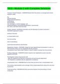
Accommodation: adjustment of the shape of the lens so light from objects at different distances
focuses correctly on retina
bring both near and far objects into focus
- how the lens adjusts its focus
o if object is far, light rays that reach the eye are parallel, brought to focus on the retina
at point A
o if object moves closer, light rays reflected from this object enter the eye at an angle
which pushed focus to point B
o light is stopped by the back of the eye before it reaches point B so image on the retina
is out of focus
o when ciliary muscles relax = lens flattens à distant objects are in focus
o ciliary muscles in the front of the eye tighten and increase curvature of the lens so that
it gets thicker
§ when lens is rounder à nearby objects are in focus
- occurs unconsciously
- has its limits
o when it is too close you cannot focus
o the distance at which lens can no longer adjust is called near point à increases with
age (presbyopia à lens hardens)
, Retina
Constriction or dilation of the pupil by the iris to control light entering eye à accommodation by the
lens to focus the light on the retina à form clear image on retina
- Retina transforms the image into neural signals to be sent to the brain
o Soft, texture tissue, curved surface, retinal image
is inverted (up = down, left = right)
- Retinal image: a clear image on the retina of the optic
array
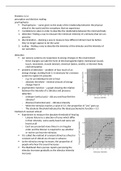
Anatomy of the retina:
- Innermost of the three membranes
- Made up of different classes of neurons with distinct
functions
Nuclear layers (contains nuclei of various types of retinal
neurons):
- Outer nuclear layer:
o Consists of photoreceptors: retinal neurons that transduce light into neural signals
à rods (greyscale, sensitive to light à responsible for dark vision) à cones (colour)
à responsible for perception and transduction
- Inner nuclear layer contains different neurons
o bipolar cells: involved in the transduction of electrical signals to ganglion cells
o horizontal cells: They help integrate and regulate the input from multiple
photoreceptor cells. Inhibit neighbouring cells. Are weakened signals
o amacrine cells:
- ganglion cell layer: pass the cells from bipolar cells to the optical layer
Source: Goldstein chapter 2 and 3,
Learning Goals:
• what is the structure of the eye and what are its functions?
• what parts of the eye play a role in perceiving light?
• What conditions/ diseases/ problems are there?
• how do different stimuli trigger our sensory stimuli? how do we perceive different grey
tones?
• How does the variation of light play a role in how we perceive things?
THE VISUAL SYSTEM
Yantis and Abrams Chapter 2
1. Light reflected from objects in the environment and enters the eye through the pupil
2. Focused by the cornea and lens to form sharp images of the objects on the retina which
contains receptors for vision
Shapes and Sizes:
- Spherical à easy to rotate and move
- Optic axis à imaginary diameter line from front to the back
Three membranes:
, - Sclera: protective Outer membrane, made up of (tough protective
covering whose visible portion in the white of eye and transparent
in front of eye)
- Choroid: Middle membrane, lines interior of the sclera, contains
blood vessels that supply inside of eye with oxygen and nutrients
- Retina: inner membrane, made up of neurons, light sensitive
receptors (convert light into neural signals) and other neurons
Cornea: transparent membrane in front of eye
- Light enters the eye by first passing through cornea which
refracts the light
- Focusing process
- Cannot adjust how much light passing through is refracted
Iris: coloured part of eye, muscle with opening, controls size of pupil by contracting and relaxing in
response to intensity of light entering
Pupil: opening through which light enters, size controlled by pupillary light reflex
- Small pupil = it constricts
- Larger = it dilates
Three chambers:
- Anterior chamber: space between the cornea and the iris
o Filled with Aqueous humor
- Posterior chamber: space between lens and iris
o Filled with Aqueous humor
- Vitreous chamber: main interior portion of eye
o Filled with Vitreous humor
Fluids:
- Aqueous humor:
o clear, thin fluid (99%+ water)
- vitreous humor
o more gel like (99%+ water)
- both slightly refract light, but refraction can’t be adjusted
- intraocular pressure: pressure of the fluids in the chambers, must be greater than air
pressure to prevent eyes from collapsing
Lens and Accommodation
Light à through cornea à refracts light à light passes through pupil à into lens
- Lens: transparent structure located directly behind pupillary aperture
o refracts the light passing through the pupil so that the light focuses properly on the
retina
, o Weak lens: doesn’t refract much light, thin, flat, long focal length
o Strong lens: refracts light sharply, thick, rounded, short focal length
- focal length: distance from the lens at which the image of an object is in focus when the
object is far away from lens
o determined by the power of any lens to refract light
- Diopters: power of a lens à 1/(focal length) in meters
o Greater the power = the shorter the focal length
- As object moves closer to lens, image of object moves further away from lens à the eye
adjusts the shape of the lens to change it’s focal length
- Zonule fibres: fibres that connect the lens to the choroid, pull on lens to change its shape
- Ciliary muscles: attached to choroid, relax (stretches lens and makes it thin, flat, weak) and
contract (lens isn’t stretched as much, thicker, rounder shape) to control how the choroid
pulls on zonule fibres to change shape of lens
Accommodation: adjustment of the shape of the lens so light from objects at different distances
focuses correctly on retina
bring both near and far objects into focus
- how the lens adjusts its focus
o if object is far, light rays that reach the eye are parallel, brought to focus on the retina
at point A
o if object moves closer, light rays reflected from this object enter the eye at an angle
which pushed focus to point B
o light is stopped by the back of the eye before it reaches point B so image on the retina
is out of focus
o when ciliary muscles relax = lens flattens à distant objects are in focus
o ciliary muscles in the front of the eye tighten and increase curvature of the lens so that
it gets thicker
§ when lens is rounder à nearby objects are in focus
- occurs unconsciously
- has its limits
o when it is too close you cannot focus
o the distance at which lens can no longer adjust is called near point à increases with
age (presbyopia à lens hardens)
, Retina
Constriction or dilation of the pupil by the iris to control light entering eye à accommodation by the
lens to focus the light on the retina à form clear image on retina
- Retina transforms the image into neural signals to be sent to the brain
o Soft, texture tissue, curved surface, retinal image
is inverted (up = down, left = right)
- Retinal image: a clear image on the retina of the optic
array
Anatomy of the retina:
- Innermost of the three membranes
- Made up of different classes of neurons with distinct
functions
Nuclear layers (contains nuclei of various types of retinal
neurons):
- Outer nuclear layer:
o Consists of photoreceptors: retinal neurons that transduce light into neural signals
à rods (greyscale, sensitive to light à responsible for dark vision) à cones (colour)
à responsible for perception and transduction
- Inner nuclear layer contains different neurons
o bipolar cells: involved in the transduction of electrical signals to ganglion cells
o horizontal cells: They help integrate and regulate the input from multiple
photoreceptor cells. Inhibit neighbouring cells. Are weakened signals
o amacrine cells:
- ganglion cell layer: pass the cells from bipolar cells to the optical layer