Lecture 5
How to study the brain
Neurological diseases and cases
Methods to study the brain and its role in behaviour/cognition
• Behavioural studies
• Manipulations of brain function
• Neuroanatomy and histology
• Electrophysiology
• Imaging (MRI and PET scans)
• Computational models/brain-based devices
Case studies: HM
- Henry G. Molaison (1926-2008)
- He had surgery to try and stop his epileptic seizures, a surgical resection of medial temporal lobe,
mainly the hippocampus.
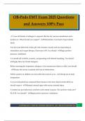
,Concepts of memory systems
- A taxonomy of mammalian memory systems:
- This taxonomy lists the brain structures and connections
thought to be especially important for each kind of declarative
and non-declarative memory.
Experimentally induced lesions and other brain manipulations
- Selective destruction of specific brain sites (mechanical, electrolytic, neurotoxic)
- Temporary pharmacological manipulations via pre-implanted micro-cannulae to switch
neurons or specific receptors on and off
- Targeted mutations of brain-specific genes
- Optogenics
- Trans-cranial magnetic stimulation (TMS)
Selective place learning deficits after hippocampal lesions in rats
Neuroanatomy study of brain connectivity
- Neuronal tract tracing
- Diffusion magnetic resonance imaging: Berg-Johansen and
Rushworth (2009)
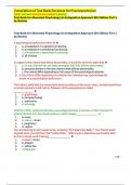
,Polymodal sensory input to the hippocampus
- Burwell (2000)
- The parahippocampal region: cortico-cortical connectivity:
Electrophysiology: Recording the electrical activity of the brain
• Single-unit recordings: recording the electrical activity of single neurons
- John O’Keefe: Nobel prize in discoveries of cells that constitute a positioning system in the brain.
- e.g. place cells in the hippocampus
• Electroencephalogram (EEG): recording electrical potentials generated by many neurons
(field potentials)
- e.g. EEG recorded from rat hippocampus
Electrophysiology in humans
• Invasive single-unit and EEG recordings
- Only conducted in rare cases for the pre-surgical evaluation of epilepsy patients (Engel et al, 2005)
, • Surface EEG
- Spontaneous and event related (evoked)
• Magnetencaphalography
- Measures the small magnetic-field changes accompanying electrical voltage changes due to
brain activity.
- Better spatial resolution than EEG (<1cm)
Magnetic Resonance Imaging (MRI)
- Images are generated from magnetic-resonance (MR) signal that emanates from hydrogen
nuclei in brain tissue when these are aligned by a strong magnetic field and then excited by a
magnetic pulse.
• Structural MRI of the brain
- Non-invasive imaging of brain structure based on MRI contrast between different tissue types due
to different densities of H nuclei
• Functional MRI of the brain
- Non-invasive imaging of brain ‘activity’ based on MR signal changes associated with metabolic and
cerebral-blood-flow changes. Most common method is based on changes in the Blood-Oxygen-Level-
Dependent (BOLD) MR signal.
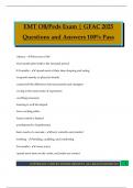
Activation of the human hippocampus during place memory task in a virtual environment:
- An fMRI study
Positron Emission Tomography (PET)
- Involves injection of radioactive tracers that resemble compounds of biological interest (e.g., 18F-2-
deoxyglucose). Using dedicated detectors around the head, these tracers can be followed in the
brain (e.g., to monitor metabolic activation).
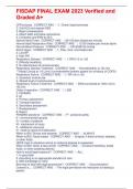
- PET imaging of brain activity and chemical neurotransmission:
• Changes in Parkinson’s
- Less DAT in striatum – reflects degeneration of dopaminergic fibres that express this transporter at
terminals
- More binding of dopamine receptor-specific tracer – reflects less dopamine
release that could displace tracer from receptor
- Some regions hypo-, others hyperactive; changes across disease course
How to study the brain
Neurological diseases and cases
Methods to study the brain and its role in behaviour/cognition
• Behavioural studies
• Manipulations of brain function
• Neuroanatomy and histology
• Electrophysiology
• Imaging (MRI and PET scans)
• Computational models/brain-based devices
Case studies: HM
- Henry G. Molaison (1926-2008)
- He had surgery to try and stop his epileptic seizures, a surgical resection of medial temporal lobe,
mainly the hippocampus.
,Concepts of memory systems
- A taxonomy of mammalian memory systems:
- This taxonomy lists the brain structures and connections
thought to be especially important for each kind of declarative
and non-declarative memory.
Experimentally induced lesions and other brain manipulations
- Selective destruction of specific brain sites (mechanical, electrolytic, neurotoxic)
- Temporary pharmacological manipulations via pre-implanted micro-cannulae to switch
neurons or specific receptors on and off
- Targeted mutations of brain-specific genes
- Optogenics
- Trans-cranial magnetic stimulation (TMS)
Selective place learning deficits after hippocampal lesions in rats
Neuroanatomy study of brain connectivity
- Neuronal tract tracing
- Diffusion magnetic resonance imaging: Berg-Johansen and
Rushworth (2009)
,Polymodal sensory input to the hippocampus
- Burwell (2000)
- The parahippocampal region: cortico-cortical connectivity:
Electrophysiology: Recording the electrical activity of the brain
• Single-unit recordings: recording the electrical activity of single neurons
- John O’Keefe: Nobel prize in discoveries of cells that constitute a positioning system in the brain.
- e.g. place cells in the hippocampus
• Electroencephalogram (EEG): recording electrical potentials generated by many neurons
(field potentials)
- e.g. EEG recorded from rat hippocampus
Electrophysiology in humans
• Invasive single-unit and EEG recordings
- Only conducted in rare cases for the pre-surgical evaluation of epilepsy patients (Engel et al, 2005)
, • Surface EEG
- Spontaneous and event related (evoked)
• Magnetencaphalography
- Measures the small magnetic-field changes accompanying electrical voltage changes due to
brain activity.
- Better spatial resolution than EEG (<1cm)
Magnetic Resonance Imaging (MRI)
- Images are generated from magnetic-resonance (MR) signal that emanates from hydrogen
nuclei in brain tissue when these are aligned by a strong magnetic field and then excited by a
magnetic pulse.
• Structural MRI of the brain
- Non-invasive imaging of brain structure based on MRI contrast between different tissue types due
to different densities of H nuclei
• Functional MRI of the brain
- Non-invasive imaging of brain ‘activity’ based on MR signal changes associated with metabolic and
cerebral-blood-flow changes. Most common method is based on changes in the Blood-Oxygen-Level-
Dependent (BOLD) MR signal.
Activation of the human hippocampus during place memory task in a virtual environment:
- An fMRI study
Positron Emission Tomography (PET)
- Involves injection of radioactive tracers that resemble compounds of biological interest (e.g., 18F-2-
deoxyglucose). Using dedicated detectors around the head, these tracers can be followed in the
brain (e.g., to monitor metabolic activation).
- PET imaging of brain activity and chemical neurotransmission:
• Changes in Parkinson’s
- Less DAT in striatum – reflects degeneration of dopaminergic fibres that express this transporter at
terminals
- More binding of dopamine receptor-specific tracer – reflects less dopamine
release that could displace tracer from receptor
- Some regions hypo-, others hyperactive; changes across disease course