NURS 201 REVIEW FOR QUIZ 3
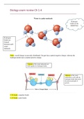
Summary NURS 201 REVIEW FOR QUIZ 3 Chapter 16 (Respiratory) Review the picture from slide 8 • Respiratory cycle: – Eupnea ▪ Regular, even, rhythmic pattern of breathing – Dyspnea ▪ Change in this pattern producing shortness of breath or difficulty breathing Slide 20,21,22,23,25, 26 (on this slide noted T3 spinous process, and 6th rib midaxillary line) (pictures slides), 27 Patient positioned and gowned for assessment- Pt. should be sitting. Pt. should only have a gown/drape. Stand in front of the pt. Lightening should be adequate to detect color differences, lesions, & chest movements. Explain the procedure & ask the pt. to breathe normally. Observe skin color & symmetry of structures. Inspect for chest configuration: The adult transverse diameter is twice that of the anteroposterior diameter (AP: T=1:2) Slide 46 (see picture) Pattern for palpating the posterior thorax- Explain that you will be palpating (touching) the pt.s back to determine if there is any area of tenderness & to inform you if pain or discomfort is felt in any area touched. Pain may occur with fibrous tissue or underlying structures: pleura. Crepitus is a crunching feeling under the skin caused by air leaking into subcutaneous tissue. Palpation for respiratory expansion- the movement of the chest during breathing by placing the hands on the lower chest & asking the pt. to take a deep breath. Place the palmar surface of your hand, with the thumbs close to the vertebrae, on the chest at the T10. Pinch up some skin between your thumbs. Ask the pt. to take & a deep breath. The movement & pressure of the chest against your hands should feel smooth & even. Your thumbs should move away from the spine & skin should move smoothly as the chest moves with inspiration. Unilateral decrease or delay in expansion may indicate underlying fibrotic or obstructive lung disease or pneumothorax. Palpation for tactile fremitus using metacarpophalangeal joint area- Fremitus is the palpable vibration on the chest wall when the client speaks (stronger over the trachea & diminishes over the bronchi & almost non-existent over the alveoli. Use one palmar surface of the hand at the base of the fingers surface; ask the pt. to repeat “ 99” or “1, 2, 3” in a clear loud voice. Decreased fremitus: soft voice, thick chest wall, obesity or underlying diseases: COPD, pleural effusion, fibrosis, or tumor. Increased fremitus occurs with fluids in the lungs or infection Slide 49. Percussion of the posterior thorax – Lungs – Diaphragmatic excursion ( She mentioned slide 49) Slide 50. There is a pattern of percussion (picture). Pattern for percussion: Posterior thorax. Place the pleximeter in the intercostal space parallel to the ribs during percussion. Standing slightly to the side of the pt. allows the pleximeter finger to lie more firmly on the chest as you move through all the thoracic areas. Percuss the apex of the left lung & then the height of the right lung. Percuss from side to side, comparing sounds. Percussion over solidified or fluid-filled areas will yield a dull sound. Percussion over bone= flat sounds. Slide 51. Diaphragmatic movement, percussion- (Diaphragmatic excursion)- It requires the use of a marker & a ruler. An asymmetric diaphragm may indicate diaphragmatic paralysis or pleural effusion of the elevated side. It involves two steps: a) Determine the level of the diaphragm during quiet respiration by placing the pleximeter finger above the expected level of diaphragmatic dullness (T7 or T8) at the mid-scapular line. Percuss in steps downward until dullness replaces resonance on both sides of the chest. Mark those areas. (T10). Asymmetric= death. Paralysis Slide 52. Diaphragmatic movement, measurement-Measure the diaphragmatic movement by asking the pt. to fully exhale. Starting at the previous markings on the left chest, percuss upward from dullness to resonance. Mark that area. Then ask the pt. to inhale fully & hold it as you begin to percuss from the level of the diaphragm downward moving from resonance to dullness. Mark that and repeat the other side. Slide 54. There is a picture for “Pattern for auscultation”. Posterior thorax- Posterior thorax- (the pattern for auscultation is the same as that for percussion). In the obese pt. the skin folds must be moved & the stethoscope placed firmly on the chest wall for auscultation. Asking the pt. to put the arm over the head & lean toward the opposite side is often helpful in accessing the chest wall during auscultation. Monitor pt.’s breathing: Prevent hyperventilation! Slide 56. • Auscultation of voice sounds – Bronchophony- As pt. says “99” normal sound should be muffled – Egophony-Ask the pt. to say “E”; in normal lung tissue you hear: “eeeeee” – The “E” sounds like; “ away” over areas of consolidation – Whispered pectoriloquy-Ask the pt. to whisper “1,2, 3”-in normal tissue the sound will be faint, almost undistinguishable, muffled Slide 64. Picture (Professor mentioned: vesicular moist loud prominent lung sound). I don’t remember what the professor means. Slide 65. Professor mentioned that tachypnea leads to alkalosis. See the below table.
Written for
- Institution
- Summary NURS 201
- Course
- Summary NURS 201
Document information
- Uploaded on
- December 9, 2022
- Number of pages
- 11
- Written in
- 2022/2023
- Type
- Exam (elaborations)
- Contains
- Questions & answers
Subjects
- even
-
summary nurs 201 review for quiz 3 chapter 16 respiratory review picture from slide 8 • respiratory cycle – eupnea ▪ regular
-
rhythmic pattern of breathing – dyspnea ▪ change in this patt