Bacteriologie
Characteristics of prokaryotic cells
Examen: 3 open vragen – max 3 kiemen gevraagd
Inleiding
Prokaryoten Eukaryoten
Algemeen - Eencellig
- Geen compartimentalisatie
- Geen histonproteïnen
DNA Circulair Lineair
Organellen - Geen membraan gebonden organellen - Wel membraan gebonden organellen
(zoals mitochondriën) - Wel nucleus
- Geen nucleus - Enkel celmembraan
- Celwand en celmembraan
- Soms flagel, fimbriae
Voorbeeld Bacteriën Parasieten en fungi
Grootte 0,5 tot 2,0 µm Humane cellen: 10 µm
Figuur
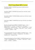
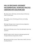
Vormen van bacteriën
- Coccen: bolletje
o Diplococcus: 2 afgeplatte bollen naast
elkaar (bv Neisseria meningitis)
o Streptococcus: coccen delen 2D
longitudinaal op een streep (bv
Streptococcus pyogenus – veroorzaakt
keelonsteking)
o Staphylococcen: coccen delen 3D (bv Staphylococcus aureus – huidinfectie)
- Bacillus: staafvorm (bv bacillus antracis – antrax)
- Streptobacillus: bacillus deelt op een streep
- Vibrio: kommavormig (bv.: Vibrio cholerae – waterige diarree)
- Spirocheet: kurkentrekkervormig (bv.: Treponema pallidum – syfillis of Borellia burgodoferie
– zieke van Lyme)
- Spirillum: medisch niet belangrijk → niet kennen
1
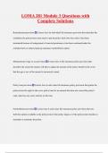
,Overzicht van structuur van bacteriën
- Celmembraan meestal omgeven door celwand
- Intern cytoplasma met ribosomen en nucleaire regio
- Variabiliteit aan externe structuren: capsule, flagellum en pilus (fimbriae)
o Bepalen of ze wel of niet herkend worden door immuunsysteem
o Bacteriën kunnen verschillen door virulentiefactoren: factoren die pathogeniciteit
bepalen en ziekte veroorzakenVoorbeeld: ene heeft dit niet bv E.Coli maakt vit K in
darmen en is niet gevaarlijk terwijl een andere E.Coli een ernstige infectie kan
veroorzaken die kan leiden tot dood → DUS 2 bacteriën met zelfde naam, maar
verschillend effect
Opbouw prokaryoten
Celwand
- Ligt rond het celmembraan
- Celmembraan: fosfolipide dubbellaag zonder sterolen (zoals cholesterol) → gaan sneller
kapot hierdoor (want sterol is nodig voor stevigheid en integriteit van membraan)
→ daarom hebben bacteriën een celwand als oplossing
- Functies:
o Vormgeving van cel
o Zorgt ervoor dat cel niet openbreekt door osmose
o Reguleert niet de entry van materialen in de cel (want is te poreus)
- Celwand bestaat uit:
o Peptidoglycanen (PG) (enkel bacteriën hebben PG = target voor GM zoals penicilline)
o Buitenste membranen
o Periplasmatische ruimte
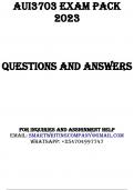
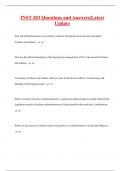
Peptidoglycanen laag = mureïne
- Disacharide backbone (polymeer): verschillende polymeren boven
elkaar om te zorgen voor de stevigheid - 2 lagen bestaan uit NAM (N-
acetylmuramic acid) en NAG (N-acetylglucosamine) die telkens worden
herhaald→ maar als disacharide polymeer alleen zou voorkomen zou
het te zwak zijn → AZ nodig
- Vorming van keten van 4 AZ (tetrapeptide) aan NAM
→ NAM gaat binden met aminogroep van EW → H2O wordt hierbij
afgesplitst
- 2 ketens zijn verbonden via een 3,4-peptidebinding
- Volgorde AZ:
1. L-alanine
2. D-glutaminezuur
3. Diaminopimelinezuur (DAP) (bij gram -) of Lysine (gram +)
4. D-alanine
2
,Waarom kan tetrapeptide enkel binden op NAM?
Want NAM heeft een carbonzuurfunctie (rood) die er voor zorgt dat een
amidinebinding kan plaatsvinden tussen NAM en L-alanine waarbij H2O vrijkomt (bij
EW gaan AZ zelfde binding aan tussen carboxyterminus en aminozuurterminus). Door
structuur van NAG gaat dat niet.
Verschil in PG tussen gram + en gram –
- Gram –
o DAP op positie 3
o Geen oligopeptidebrug tussen 3 en 4 → rechtstreekse binding tussen DAP en D-
alanine = 3,4 peptidebinding
- Gram +
o L-lysine op plaats 3
o Wel echte oligopeptidebrug (van 5 glycine moleculen) tussen L-lysine en D-alanine
Gramkleuring is gebaseerd op het verschil in celwand van de 2
Celwand van gram + bacteriën
- Dikke peptidoglycaanlaag
o Neemt 60 – 90% van celwand in
o Als er geen PG is = protoplast (=1
celmembraan)
- Teichoïnezuren
o Gewone teichoïnezuren vertrekken uit PG laag
o Lipoteichoïnezuren vertrekken uit
fosfolipidenlaag
o Opbouw
▪ Ribitol- of glycerol fosfaat
▪ Fosfaatgroep (Functie: negatief geladen – vangen van Ca2+ en Mg2+ en
transporteren)
▪ AZ en suikers – oppervlakte AG
- Eiwitten
o Adhesiefactoren
o Capsule productie
o Peniciline binding protein (PBP): zit in celmembraan - group of proteins that are
characterized by their affinity for and binding of penicillin. They are a normal
constituent of many bacteria. They are involved in the final stages of the synthesis
of peptidoglycan
- 1 fosfolipdendubbellaag
3
, Celwand van gram – bacteriën
- Dunne PG laag (10 nm)
o 10 – 20% van celwand
o Indien geen PG → sferoplast (=2
celmembranen)
- Periplasmatische ruimte:
o Tussen binnenste en buitenste
membraan
o Bestaat uit: PG laag, toxines en
katabole enzymen
- Eiwitten: adhesie factoren, capsule producite
en PBP (zelfde als gram +) in celmembraan
- Lipopolysachariden (LPS): zitten op buitenste celmembraan (zoals soort antennes)
- 2 celmebranen (2 fosfolipidendubbellagen met daartussen PG)
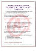
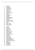
LPS opbouw:
1. Polysacharide
o Core oligosacharide: 10 – 15 heptose (7C) en octose (8C) suikers (KDO = keto-
deoxyoctulosonaat
o Somatisch AG = O-antigen repeat: hexose (6C) repeat
▪ Grootste variatie tussen bacteriële soorten
▪ Belangrijk voor classificaite
2. Lipide A: endotoxine (=toxisch voor mensen en dieren-
o Gefosforyleerd diglucosamine: diglucosamine (2G) omgeven door 2 fosfaatgroepen:
P-G-G-P-G-G-P-…
→ relatief weinig variatie hierop
o Meerdere C14 vetzuren op de G (zoals myristinezuur C14) → gevaarlijk als het
intraveneus wordt toegediend
LPS functie:
1. Aanhechting aan verschillende weefsels
2. AG variatie (is een soort AG)
3. Beschermende barrière: negatief geladen fosfaatgroepen tussen
2 LPS moleculen in het membraan stoten elkaar normaal af →
Ca2+ gaat binden en zorgt voor stabilisatie van LPS molecule →
Antibioticum kan hier dan moeilijker door
→ MAAR EDTA (chelator) is ook actieve stof van veel AB → gaat
Ca2+ wegnemen → geen barrière meer
4
Characteristics of prokaryotic cells
Examen: 3 open vragen – max 3 kiemen gevraagd
Inleiding
Prokaryoten Eukaryoten
Algemeen - Eencellig
- Geen compartimentalisatie
- Geen histonproteïnen
DNA Circulair Lineair
Organellen - Geen membraan gebonden organellen - Wel membraan gebonden organellen
(zoals mitochondriën) - Wel nucleus
- Geen nucleus - Enkel celmembraan
- Celwand en celmembraan
- Soms flagel, fimbriae
Voorbeeld Bacteriën Parasieten en fungi
Grootte 0,5 tot 2,0 µm Humane cellen: 10 µm
Figuur
Vormen van bacteriën
- Coccen: bolletje
o Diplococcus: 2 afgeplatte bollen naast
elkaar (bv Neisseria meningitis)
o Streptococcus: coccen delen 2D
longitudinaal op een streep (bv
Streptococcus pyogenus – veroorzaakt
keelonsteking)
o Staphylococcen: coccen delen 3D (bv Staphylococcus aureus – huidinfectie)
- Bacillus: staafvorm (bv bacillus antracis – antrax)
- Streptobacillus: bacillus deelt op een streep
- Vibrio: kommavormig (bv.: Vibrio cholerae – waterige diarree)
- Spirocheet: kurkentrekkervormig (bv.: Treponema pallidum – syfillis of Borellia burgodoferie
– zieke van Lyme)
- Spirillum: medisch niet belangrijk → niet kennen
1
,Overzicht van structuur van bacteriën
- Celmembraan meestal omgeven door celwand
- Intern cytoplasma met ribosomen en nucleaire regio
- Variabiliteit aan externe structuren: capsule, flagellum en pilus (fimbriae)
o Bepalen of ze wel of niet herkend worden door immuunsysteem
o Bacteriën kunnen verschillen door virulentiefactoren: factoren die pathogeniciteit
bepalen en ziekte veroorzakenVoorbeeld: ene heeft dit niet bv E.Coli maakt vit K in
darmen en is niet gevaarlijk terwijl een andere E.Coli een ernstige infectie kan
veroorzaken die kan leiden tot dood → DUS 2 bacteriën met zelfde naam, maar
verschillend effect
Opbouw prokaryoten
Celwand
- Ligt rond het celmembraan
- Celmembraan: fosfolipide dubbellaag zonder sterolen (zoals cholesterol) → gaan sneller
kapot hierdoor (want sterol is nodig voor stevigheid en integriteit van membraan)
→ daarom hebben bacteriën een celwand als oplossing
- Functies:
o Vormgeving van cel
o Zorgt ervoor dat cel niet openbreekt door osmose
o Reguleert niet de entry van materialen in de cel (want is te poreus)
- Celwand bestaat uit:
o Peptidoglycanen (PG) (enkel bacteriën hebben PG = target voor GM zoals penicilline)
o Buitenste membranen
o Periplasmatische ruimte
Peptidoglycanen laag = mureïne
- Disacharide backbone (polymeer): verschillende polymeren boven
elkaar om te zorgen voor de stevigheid - 2 lagen bestaan uit NAM (N-
acetylmuramic acid) en NAG (N-acetylglucosamine) die telkens worden
herhaald→ maar als disacharide polymeer alleen zou voorkomen zou
het te zwak zijn → AZ nodig
- Vorming van keten van 4 AZ (tetrapeptide) aan NAM
→ NAM gaat binden met aminogroep van EW → H2O wordt hierbij
afgesplitst
- 2 ketens zijn verbonden via een 3,4-peptidebinding
- Volgorde AZ:
1. L-alanine
2. D-glutaminezuur
3. Diaminopimelinezuur (DAP) (bij gram -) of Lysine (gram +)
4. D-alanine
2
,Waarom kan tetrapeptide enkel binden op NAM?
Want NAM heeft een carbonzuurfunctie (rood) die er voor zorgt dat een
amidinebinding kan plaatsvinden tussen NAM en L-alanine waarbij H2O vrijkomt (bij
EW gaan AZ zelfde binding aan tussen carboxyterminus en aminozuurterminus). Door
structuur van NAG gaat dat niet.
Verschil in PG tussen gram + en gram –
- Gram –
o DAP op positie 3
o Geen oligopeptidebrug tussen 3 en 4 → rechtstreekse binding tussen DAP en D-
alanine = 3,4 peptidebinding
- Gram +
o L-lysine op plaats 3
o Wel echte oligopeptidebrug (van 5 glycine moleculen) tussen L-lysine en D-alanine
Gramkleuring is gebaseerd op het verschil in celwand van de 2
Celwand van gram + bacteriën
- Dikke peptidoglycaanlaag
o Neemt 60 – 90% van celwand in
o Als er geen PG is = protoplast (=1
celmembraan)
- Teichoïnezuren
o Gewone teichoïnezuren vertrekken uit PG laag
o Lipoteichoïnezuren vertrekken uit
fosfolipidenlaag
o Opbouw
▪ Ribitol- of glycerol fosfaat
▪ Fosfaatgroep (Functie: negatief geladen – vangen van Ca2+ en Mg2+ en
transporteren)
▪ AZ en suikers – oppervlakte AG
- Eiwitten
o Adhesiefactoren
o Capsule productie
o Peniciline binding protein (PBP): zit in celmembraan - group of proteins that are
characterized by their affinity for and binding of penicillin. They are a normal
constituent of many bacteria. They are involved in the final stages of the synthesis
of peptidoglycan
- 1 fosfolipdendubbellaag
3
, Celwand van gram – bacteriën
- Dunne PG laag (10 nm)
o 10 – 20% van celwand
o Indien geen PG → sferoplast (=2
celmembranen)
- Periplasmatische ruimte:
o Tussen binnenste en buitenste
membraan
o Bestaat uit: PG laag, toxines en
katabole enzymen
- Eiwitten: adhesie factoren, capsule producite
en PBP (zelfde als gram +) in celmembraan
- Lipopolysachariden (LPS): zitten op buitenste celmembraan (zoals soort antennes)
- 2 celmebranen (2 fosfolipidendubbellagen met daartussen PG)
LPS opbouw:
1. Polysacharide
o Core oligosacharide: 10 – 15 heptose (7C) en octose (8C) suikers (KDO = keto-
deoxyoctulosonaat
o Somatisch AG = O-antigen repeat: hexose (6C) repeat
▪ Grootste variatie tussen bacteriële soorten
▪ Belangrijk voor classificaite
2. Lipide A: endotoxine (=toxisch voor mensen en dieren-
o Gefosforyleerd diglucosamine: diglucosamine (2G) omgeven door 2 fosfaatgroepen:
P-G-G-P-G-G-P-…
→ relatief weinig variatie hierop
o Meerdere C14 vetzuren op de G (zoals myristinezuur C14) → gevaarlijk als het
intraveneus wordt toegediend
LPS functie:
1. Aanhechting aan verschillende weefsels
2. AG variatie (is een soort AG)
3. Beschermende barrière: negatief geladen fosfaatgroepen tussen
2 LPS moleculen in het membraan stoten elkaar normaal af →
Ca2+ gaat binden en zorgt voor stabilisatie van LPS molecule →
Antibioticum kan hier dan moeilijker door
→ MAAR EDTA (chelator) is ook actieve stof van veel AB → gaat
Ca2+ wegnemen → geen barrière meer
4