Cytology - study of cells.
either light microscopes or electron
Cells can be studied in detail with microscopes,
microscopes.
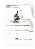
Label the diagram of a light microscope;
-
eyepiece
corse focus
nose piece
objective
Fine focus lenses
x 4 x 10 X 40
stage
stage clip
diaphram lever-controls amount of light through
condenser
stage -light source/camp
height adjustment
Magnification = how many times bigger
the image is when
compared to the object
atoms + molecules
f
Maximum magnification LM = 1500 x
100,000
SEM= X TEM=
x 500,000
if use blue
light *300,000
↑biological structures
Resolution = the minimum distance apart that two objects can be in order for them to
appear as separate items.
Maximum resolution 200 nm
SEM= LOnm
0.2 TEM= 0.1 nm apart
rem
either light microscopes or electron
Cells can be studied in detail with microscopes,
microscopes.
Label the diagram of a light microscope;
-
eyepiece
corse focus
nose piece
objective
Fine focus lenses
x 4 x 10 X 40
stage
stage clip
diaphram lever-controls amount of light through
condenser
stage -light source/camp
height adjustment
Magnification = how many times bigger
the image is when
compared to the object
atoms + molecules
f
Maximum magnification LM = 1500 x
100,000
SEM= X TEM=
x 500,000
if use blue
light *300,000
↑biological structures
Resolution = the minimum distance apart that two objects can be in order for them to
appear as separate items.
Maximum resolution 200 nm
SEM= LOnm
0.2 TEM= 0.1 nm apart
rem