Neurocognition lecture 1: The brain and cognition over the life span
Brain structure and anatomy
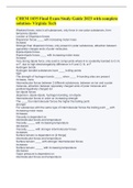
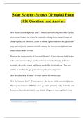
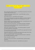
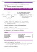
Structures (neurons):
Cell body
Axon
Axon hillock
Dendrites
Synapse
Myelin sheath
Types of neurons:
- Sensory (afferent (towards the brain)
- Interneurons (stellate, pyramidal, purkinje)
- Motor (efferent (away from the brain)
Action potentials:
Thresholded, non-decremental, all or nothing response
Triggered by summation of excitatory potentials
Driven by varying ion permeability of cell membrane
Can travel for a meter or more
Triggers neurotransmitter release at axon terminal
Synapse
Neurotransmitters:
Acetylcholine, dopamine, norepinephrine, serotonin, glutamine, gamma-aminobutyric acid
(GABA)
Receptor cells in the postsynaptic membrane van adapt to under or over-use
The distribution of synapses connecting to a cell influences its excitability.
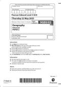
Glia cells:
- Astrocytes (Blood-brain barrier, structural support)
- Oligodendrocytes - Myelin for CNS neurons
- Microglial cells - Fight infections, waste disposal –
Ependymal cells - Ventricular surface epithelium, create CSF
Schwann cells - Myelin for peripheral neurons
Cortical cell layers:
Different types of neuron are often organized in layers
Sensory (input), interneurons (relay) and motor (output) neurons are grouped
Layers are different in different cortical areas, depending on primary function
Each layer has a specific function.
White matter tracts (because of the myelin)
Bundles of myelinated axons. Connection neurons throughout the central and peripheral
nervous system
Types of fibers:
- Association fibers connecting areas within a hemisphere
,- Commissural fibers crossing to the other hemisphere, to the same (homotopic) or a different
place (heterotopic)
- Projection fibers connect outward, to subcortical regions, cerebellum or the spinal chord
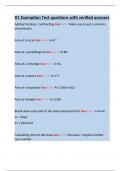
Major component of the CNS
- Forebrain (incl. hemispheres, corpus callosum and subcortical deep structure
(telencephalon)
- Diencephalon (incl. thalamic structures)
- Midbrain (mesencephalon) (top of the brain stem, incl. sensory and motor relay
nuclei)
- Hindbrain (metencephalon) (incl. pons and cerebellum, medulla oblongata)
- Spinal chord
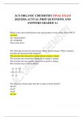
Hindbrain and midbrain
- Medulla oblongata
- Pons
- Cerebellum
- Origin of cranial nerves v-xii
Midbrain
- Superior/inferior colliculi
- Substantia nigra
- Origin of cranial nerves
Diencephalon
- Thalamus, hypothalamus, pituary gland
Diencephalon (thalamus)
Telencephalon or cerebrum (subcortical = anything below/surrounded by the cortex)
- Basal ganglia (motor, associative, reward circuit)
- Limbic structures (stress response, emotional processing)
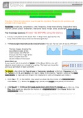
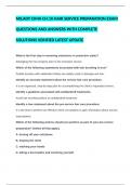
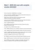
Telencephalon (cortical)
- Frontal lobes: movement, attention, reward, short-term memory, planning, impulse
control, and more
- Parietal lobes: sensory integration, association processes, language functions, spatial
processing, sense of touch, some visual processes, and more
- Occipital lobes: mainly primary visual areas
- Temporal lobes: memory, emotion association, primary auditory areas, some visual
processes, and more!
Lateralization: symmetry and asymmetry
Asymmetries:
- Language (left)
- Global perception (right)
- Creative vs logical
Gyri and culci
- Gyrus (bumb)
- Sulcus (groove)
are recognizable landmarks
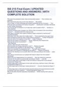
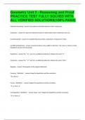
,Ventricles of the brain
- Lateral
- 3rd
- Aqueduct
- 4th
- Central
CSF (fluid) runs through the ventricles, subarachnoid space and the venous sinus)
Created in the lateral ventricles from arterial blood in the choroid plexus and ependymal cells)
Meninges
Membranes that cover the brain and spinal chord from the outside:
- Dura mater
- Arachnoid with subarachnoid space (CSF)
- Pia mater
Different types of naming:
- Brodmann areas (histological, types of cells)
- Functional names
- Relative locations
- Coordinate systems
Directional planes
Coordinate systems
Getting oriented in the brain (x, y and x coordicates)
Brain development and plasticity
Cell development:
- Dendritic spine formation (based on stimulation levels, connectivity between cells can
change) (dependent on life stage, long-term potentiation or depression can influence
formation or pruning of receptive spines on the dendrites)
, - Neurogenesis (contrary to long-held beliefs, some brain areas are now known to be
able to grow new neurons)
- Apoptosis (different from necrosis (cell death due to external causes), neurons van
also self-initiate preprogrammed cell death, also called pruning)
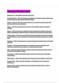
Brain structure changes with healthy aging:
Structural:
- Cortical thinning/atrophy
- Neuronal loss and loss of synapses
- White matter lesions
- Inflammations
- Decrease in cerebral blood flow
- Beta-amyloid plaques, neurofibrillary tangles degradation of cells
- Specific regions: temporal, subcortical, hippocampus etc.
Functional:
- Increased activation for same tasks (reorganization and compensation)
- Different activation
Brain damage: impact on neural structure
- Normal aging (atrophy)
- Vascular (stroke) (ischemic = lack of oxygen, blockage) (hemorrhagic = leak in the
vascular system)
- Trauma (impact) (depends on place of the damage)
- Tumors (it pushes tissue out of the way)
- Developmental disorders, some hereditary (including neurodegenerative disorders)
- Toxicity
- Infections
Development and cognition
As we age, various cognitive abilities tend to worsen:
Cognition
- Processing/psychomotor speed lower
- Working memory lower
- Episodic memory lower
- Verbal abilities no change/higher (more vocabulary)
Predictors of cognitive decline
- Age
- Medical health/biomarkers (comorbidity, genetic predisposition, HPA axis
dysregulation, neural markers)
- Experience, lifestyle, etc.
Brain reserve (anatomical differences between people)
Cognitive reserve (differences in brain function that could explain differences in susceptibility
to functional impairment in the presence of pathology)
Brain structure and anatomy
Structures (neurons):
Cell body
Axon
Axon hillock
Dendrites
Synapse
Myelin sheath
Types of neurons:
- Sensory (afferent (towards the brain)
- Interneurons (stellate, pyramidal, purkinje)
- Motor (efferent (away from the brain)
Action potentials:
Thresholded, non-decremental, all or nothing response
Triggered by summation of excitatory potentials
Driven by varying ion permeability of cell membrane
Can travel for a meter or more
Triggers neurotransmitter release at axon terminal
Synapse
Neurotransmitters:
Acetylcholine, dopamine, norepinephrine, serotonin, glutamine, gamma-aminobutyric acid
(GABA)
Receptor cells in the postsynaptic membrane van adapt to under or over-use
The distribution of synapses connecting to a cell influences its excitability.
Glia cells:
- Astrocytes (Blood-brain barrier, structural support)
- Oligodendrocytes - Myelin for CNS neurons
- Microglial cells - Fight infections, waste disposal –
Ependymal cells - Ventricular surface epithelium, create CSF
Schwann cells - Myelin for peripheral neurons
Cortical cell layers:
Different types of neuron are often organized in layers
Sensory (input), interneurons (relay) and motor (output) neurons are grouped
Layers are different in different cortical areas, depending on primary function
Each layer has a specific function.
White matter tracts (because of the myelin)
Bundles of myelinated axons. Connection neurons throughout the central and peripheral
nervous system
Types of fibers:
- Association fibers connecting areas within a hemisphere
,- Commissural fibers crossing to the other hemisphere, to the same (homotopic) or a different
place (heterotopic)
- Projection fibers connect outward, to subcortical regions, cerebellum or the spinal chord
Major component of the CNS
- Forebrain (incl. hemispheres, corpus callosum and subcortical deep structure
(telencephalon)
- Diencephalon (incl. thalamic structures)
- Midbrain (mesencephalon) (top of the brain stem, incl. sensory and motor relay
nuclei)
- Hindbrain (metencephalon) (incl. pons and cerebellum, medulla oblongata)
- Spinal chord
Hindbrain and midbrain
- Medulla oblongata
- Pons
- Cerebellum
- Origin of cranial nerves v-xii
Midbrain
- Superior/inferior colliculi
- Substantia nigra
- Origin of cranial nerves
Diencephalon
- Thalamus, hypothalamus, pituary gland
Diencephalon (thalamus)
Telencephalon or cerebrum (subcortical = anything below/surrounded by the cortex)
- Basal ganglia (motor, associative, reward circuit)
- Limbic structures (stress response, emotional processing)
Telencephalon (cortical)
- Frontal lobes: movement, attention, reward, short-term memory, planning, impulse
control, and more
- Parietal lobes: sensory integration, association processes, language functions, spatial
processing, sense of touch, some visual processes, and more
- Occipital lobes: mainly primary visual areas
- Temporal lobes: memory, emotion association, primary auditory areas, some visual
processes, and more!
Lateralization: symmetry and asymmetry
Asymmetries:
- Language (left)
- Global perception (right)
- Creative vs logical
Gyri and culci
- Gyrus (bumb)
- Sulcus (groove)
are recognizable landmarks
,Ventricles of the brain
- Lateral
- 3rd
- Aqueduct
- 4th
- Central
CSF (fluid) runs through the ventricles, subarachnoid space and the venous sinus)
Created in the lateral ventricles from arterial blood in the choroid plexus and ependymal cells)
Meninges
Membranes that cover the brain and spinal chord from the outside:
- Dura mater
- Arachnoid with subarachnoid space (CSF)
- Pia mater
Different types of naming:
- Brodmann areas (histological, types of cells)
- Functional names
- Relative locations
- Coordinate systems
Directional planes
Coordinate systems
Getting oriented in the brain (x, y and x coordicates)
Brain development and plasticity
Cell development:
- Dendritic spine formation (based on stimulation levels, connectivity between cells can
change) (dependent on life stage, long-term potentiation or depression can influence
formation or pruning of receptive spines on the dendrites)
, - Neurogenesis (contrary to long-held beliefs, some brain areas are now known to be
able to grow new neurons)
- Apoptosis (different from necrosis (cell death due to external causes), neurons van
also self-initiate preprogrammed cell death, also called pruning)
Brain structure changes with healthy aging:
Structural:
- Cortical thinning/atrophy
- Neuronal loss and loss of synapses
- White matter lesions
- Inflammations
- Decrease in cerebral blood flow
- Beta-amyloid plaques, neurofibrillary tangles degradation of cells
- Specific regions: temporal, subcortical, hippocampus etc.
Functional:
- Increased activation for same tasks (reorganization and compensation)
- Different activation
Brain damage: impact on neural structure
- Normal aging (atrophy)
- Vascular (stroke) (ischemic = lack of oxygen, blockage) (hemorrhagic = leak in the
vascular system)
- Trauma (impact) (depends on place of the damage)
- Tumors (it pushes tissue out of the way)
- Developmental disorders, some hereditary (including neurodegenerative disorders)
- Toxicity
- Infections
Development and cognition
As we age, various cognitive abilities tend to worsen:
Cognition
- Processing/psychomotor speed lower
- Working memory lower
- Episodic memory lower
- Verbal abilities no change/higher (more vocabulary)
Predictors of cognitive decline
- Age
- Medical health/biomarkers (comorbidity, genetic predisposition, HPA axis
dysregulation, neural markers)
- Experience, lifestyle, etc.
Brain reserve (anatomical differences between people)
Cognitive reserve (differences in brain function that could explain differences in susceptibility
to functional impairment in the presence of pathology)