Introduction to microscopy
Wednesday, 20 April 2022 13:28
Visible Light
• 400nm-700nm
Resolving power of the eye (limit)
• Rpeye = 0.1mm
• i.e. beyond this is invisible to our eyes
Simple lens Compound microscope
• Limit : < 6-10 fold magnification
Two-stage magnification : objective & eyepiece
Objective - magnifies + focuses; forms primary image
Eyepiece - magnified virtual image on retina of eye
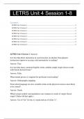
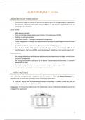
Monocular vs Binocular Microscopes
Carry microscope upright (prevents eyepieces from falling out)
Condenser • Focus light onto the specimen
• Match the NA of objective
Objective lens • Oil immersion (high power objective-usually 100X), oil medium to focus light
• Parfocal i.e. microscope that stays approx. in focus when magnification changes
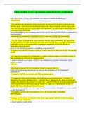
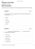
Numerical Aperture
• ↑ NA = ↑ RESOLUTION
• n-refractive index of medium between specimen and the objective
• µ-half cone of light entering the objective
• N.A. = n sin µ
Cell biology Page 1
, • ↑ NA = ↑ RESOLUTION
• n-refractive index of medium between specimen and the objective
• µ-half cone of light entering the objective
• N.A. = n sin µ
N.A. max = 1.4 (oil objective)
R = 0.61λ/N.A; increase resolution use light with shortest wavelength
Field of view
Area seen through a lens and varies with magnification
Total magnification = Mobjective × Meyepiece
Steps in tissue processing
Fixation Preserve structure to state resembling original living state; avoid artifacts
Dehydration Ethanol and acetone (e.g.) ; gradual increase in []; water content reduced
Infiltration Xylene and paraffin wax
Embedding Supports thin sectioning e.g. paraffin wax
Sectioning Provides thin sections for examinations, collected onto glass slides from water
Staining Visual contrast to identify tissue components
Why do we stain sections?
• Cells are transparent
• Stain confer contrast thus making tissue visible
• Identify structures
Basophilic Acidophilic
• Basic stained structures • Acid stained structures
• e.g. chromatin, ribosomes • e.g. collagen fibres, RBC, muscle filaments, mitochondria
• Turn red/pink (H&E)
• Hematoxylin (basic dye) stains acidic components of cells blue (basophilia)
• Eosin (acidic dye) stains basic components of cells pink (acidphilia)
Artifacts
-alters natural appearance of cells
Precursors
• Fixation artifacts
• Cell shrink or swell
• Compression or stretching=extracellular space distorted
• Scores, ripples, wrinkles
Cell biology Page 2
, Introduction to histology
Wednesday, 20 April 2022 08:53
Introduction to histology
Tuesday, 19 April 2022
14:38
Tissues = cells + extracellular matrix (mutually dependent)
ECM-mechanical structural support, transports nutrients to cell ,carry metabolised and secretory products away from cell
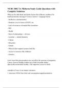
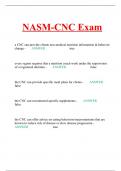
Types of Cells
Nerve Blood Muscle Fat
Muscles Tissue
• Muscle cells = muscles tissues (active contractile tissue)
• Function-produce force, cause motion (locomotion/movement within internal organs)
Description Function Location
Smooth Spindle-shaped cells with Propels substances or objects along Walls of hollow
muscle central nuclei, no internal passageways, involuntary control organs
(visceral) striations, cells arranged
closely to form sheets
Skeletal Long, cylindrical, Voluntary movement, locomotion, Attached to bones
muscle multinucleate cells, manipulation of enviro. , facial expression, or occasionally skin
obvious striations voluntary control
Cardiac Branching, striated, As it contracts, it propels blood into the Walls of heart
muscle generally uninucleate cells circulation; involuntary control
Neural Tissue
• CNS (brain & spinal cord) + peripheral nervous system (cranial nerves, spinal nerves)
• Function: transmit electrical signals from sensory receptors to effectors
• Location: brain, spinal cord and nerves
Epithelia
Cell biology Page 3
Wednesday, 20 April 2022 13:28
Visible Light
• 400nm-700nm
Resolving power of the eye (limit)
• Rpeye = 0.1mm
• i.e. beyond this is invisible to our eyes
Simple lens Compound microscope
• Limit : < 6-10 fold magnification
Two-stage magnification : objective & eyepiece
Objective - magnifies + focuses; forms primary image
Eyepiece - magnified virtual image on retina of eye
Monocular vs Binocular Microscopes
Carry microscope upright (prevents eyepieces from falling out)
Condenser • Focus light onto the specimen
• Match the NA of objective
Objective lens • Oil immersion (high power objective-usually 100X), oil medium to focus light
• Parfocal i.e. microscope that stays approx. in focus when magnification changes
Numerical Aperture
• ↑ NA = ↑ RESOLUTION
• n-refractive index of medium between specimen and the objective
• µ-half cone of light entering the objective
• N.A. = n sin µ
Cell biology Page 1
, • ↑ NA = ↑ RESOLUTION
• n-refractive index of medium between specimen and the objective
• µ-half cone of light entering the objective
• N.A. = n sin µ
N.A. max = 1.4 (oil objective)
R = 0.61λ/N.A; increase resolution use light with shortest wavelength
Field of view
Area seen through a lens and varies with magnification
Total magnification = Mobjective × Meyepiece
Steps in tissue processing
Fixation Preserve structure to state resembling original living state; avoid artifacts
Dehydration Ethanol and acetone (e.g.) ; gradual increase in []; water content reduced
Infiltration Xylene and paraffin wax
Embedding Supports thin sectioning e.g. paraffin wax
Sectioning Provides thin sections for examinations, collected onto glass slides from water
Staining Visual contrast to identify tissue components
Why do we stain sections?
• Cells are transparent
• Stain confer contrast thus making tissue visible
• Identify structures
Basophilic Acidophilic
• Basic stained structures • Acid stained structures
• e.g. chromatin, ribosomes • e.g. collagen fibres, RBC, muscle filaments, mitochondria
• Turn red/pink (H&E)
• Hematoxylin (basic dye) stains acidic components of cells blue (basophilia)
• Eosin (acidic dye) stains basic components of cells pink (acidphilia)
Artifacts
-alters natural appearance of cells
Precursors
• Fixation artifacts
• Cell shrink or swell
• Compression or stretching=extracellular space distorted
• Scores, ripples, wrinkles
Cell biology Page 2
, Introduction to histology
Wednesday, 20 April 2022 08:53
Introduction to histology
Tuesday, 19 April 2022
14:38
Tissues = cells + extracellular matrix (mutually dependent)
ECM-mechanical structural support, transports nutrients to cell ,carry metabolised and secretory products away from cell
Types of Cells
Nerve Blood Muscle Fat
Muscles Tissue
• Muscle cells = muscles tissues (active contractile tissue)
• Function-produce force, cause motion (locomotion/movement within internal organs)
Description Function Location
Smooth Spindle-shaped cells with Propels substances or objects along Walls of hollow
muscle central nuclei, no internal passageways, involuntary control organs
(visceral) striations, cells arranged
closely to form sheets
Skeletal Long, cylindrical, Voluntary movement, locomotion, Attached to bones
muscle multinucleate cells, manipulation of enviro. , facial expression, or occasionally skin
obvious striations voluntary control
Cardiac Branching, striated, As it contracts, it propels blood into the Walls of heart
muscle generally uninucleate cells circulation; involuntary control
Neural Tissue
• CNS (brain & spinal cord) + peripheral nervous system (cranial nerves, spinal nerves)
• Function: transmit electrical signals from sensory receptors to effectors
• Location: brain, spinal cord and nerves
Epithelia
Cell biology Page 3