1.4 HUMAN BODY
Lecture 1 (nervous system and brain anatomy)
The body uses two systems for communication
a) nervous system: consists of the brain, spine and nerves. It can be divided anatomically in
a central (brain and spine) part and the peripheral part (nerves and roots to the limbs).
The pheriperal nervous system can be divided by function. The somatic NS regulates
Interaction with external environment. The autonomic NS regulates the body’s internal
environment.. This can be divided in sympathetic NS (psychological arousal;; uses energy)
and parasympathetic (psychological relaxation or conservation of energy). Each part of
the body is influences by both the sympathetic and parasympathetic NS.
The central nervous system and the peripheral nervous system are made out of two types
of cells
1. Neurons: specialized in receiving information and passing it to other cells.
2. Glial cells: they don’t receive information. There are more of this cells than
neurons in the brain.
Features of a typical neuron
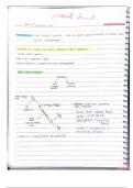
Dendrites: receive information for other neurons. It contains receptors.
Cell body: center of the neuron
Nucleus
Myelin sheath: isolation material that covers the axon and speeds up the information
transmitted
Axon: specialized in transmission
Terminal buttons: where the chemicals are being released. They firm synapses with
another neuron, its where they interact between each other.
3 types of neurons
Sensory neurons: from senses to brain. The cell body is in the middle of the neuron
instead of the end like a normal neuron. The sensory endings are located in the skin and
the information received is being passed to the soma and then to the beginning of the
neuron.
Motor neurons: from brain to muscles. It looks like a typical neuron described before.
Interneurons: send messages between neurons
The neurons differ also in the direction in which they send their information. The afferent
neurons bring information into a structure. Thus, the sensory neurons are afferent neurons. An
efferent neuron sends information away from a structure. For example, motor neurons. The
same motor neurons are also afferent. The brain is part of the nervous systems. It consists of
many brain structures that have different functions. The division of the brain during 3-4 weeks of
embryo
, Forebrain: it later develops into the telecephalon (contains the cereberum) and the
diencephalon (contains the eye cup, thalamus, hypothalamus, and epithalamus). Most
prominent part of the brain
o Cerebelum or telecephalon: consists of two sides of the brain. The two cerebral
hemispheres consist of cerebral cortex and inner structures.
o Thalamus or diencephalon: is a relay station, it sends incoming sensory
information to the cortex
Midbrain: develops into the mesencephalon (contains the midbrain)
o Tectum: superior collicullus (vision) and inferior colliculus (hearing)
o Tegmentum: several sets of nuclei and one important one is substantia nigra
(movement).
Hindbrain: develops into the myelencephalon (contains the pons and cerebellum) and
metencephalon (contains the medulla oblongata).
o Cerebellum: coordination of movement and also in cognition
o Pons: plays a role in sleeping/waking/dreaming and transmission of information
between the cortex and cerebellum
o Medulla: contains reticular formation, involved with breathing, swallowing, blood
circulation, secretion of saliva, amongst other.
We can examine the brain in three different ways
Horizontal: cut it in the middle to see the up (dorsal) and down (ventral) of the brain
Sagital: the brain is cut in the middle dividing the left and right hemisphere. We can see
the corpus callosum which connects the tract between the hemispheres. The anterior and
posterior commissures are smaller connecting tracts. Ipsilateral is when a part is on only
one of the hemispheres and contralateral is when the part is in both of the hemispheres.
Medial is when the part is in the middle and lateral is when the part is more at a side
Coronal: cuts the brain in half to see the front (rostral) and back (caudal)..
The cerebral cortex contains many cell bodies (soma of the neuron) of gray matter. It has sulci
(furrows or valleys in the cerebral cortex) and gyri (ridges or hills between the furrows).
Underneath the cerebral cortex has a white matter because the axons of neurons are covered
with myelin. There is also subcortical structures (nuclei) and cavities. Some subcortical structures
include the amygdala, hypocampus, nucleus accumbent and others. The cerebral cortex can be
divided in 4 lobes
Frontal lobe. The central sulcus is the one that makes the difference between the parietal
lobe and the frontal lobe. The high cognitive functions are contained here and motor
control also.
Temporal lobe: involved in hearing, playing an important role in language and memory.
Parietal lobe: involved in processing information from our body; sensory information.
Also involved in processing spacial information (space where we are moving).
Occipital lobe: involved in many aspects of vision.
Lecture 1 (nervous system and brain anatomy)
The body uses two systems for communication
a) nervous system: consists of the brain, spine and nerves. It can be divided anatomically in
a central (brain and spine) part and the peripheral part (nerves and roots to the limbs).
The pheriperal nervous system can be divided by function. The somatic NS regulates
Interaction with external environment. The autonomic NS regulates the body’s internal
environment.. This can be divided in sympathetic NS (psychological arousal;; uses energy)
and parasympathetic (psychological relaxation or conservation of energy). Each part of
the body is influences by both the sympathetic and parasympathetic NS.
The central nervous system and the peripheral nervous system are made out of two types
of cells
1. Neurons: specialized in receiving information and passing it to other cells.
2. Glial cells: they don’t receive information. There are more of this cells than
neurons in the brain.
Features of a typical neuron
Dendrites: receive information for other neurons. It contains receptors.
Cell body: center of the neuron
Nucleus
Myelin sheath: isolation material that covers the axon and speeds up the information
transmitted
Axon: specialized in transmission
Terminal buttons: where the chemicals are being released. They firm synapses with
another neuron, its where they interact between each other.
3 types of neurons
Sensory neurons: from senses to brain. The cell body is in the middle of the neuron
instead of the end like a normal neuron. The sensory endings are located in the skin and
the information received is being passed to the soma and then to the beginning of the
neuron.
Motor neurons: from brain to muscles. It looks like a typical neuron described before.
Interneurons: send messages between neurons
The neurons differ also in the direction in which they send their information. The afferent
neurons bring information into a structure. Thus, the sensory neurons are afferent neurons. An
efferent neuron sends information away from a structure. For example, motor neurons. The
same motor neurons are also afferent. The brain is part of the nervous systems. It consists of
many brain structures that have different functions. The division of the brain during 3-4 weeks of
embryo
, Forebrain: it later develops into the telecephalon (contains the cereberum) and the
diencephalon (contains the eye cup, thalamus, hypothalamus, and epithalamus). Most
prominent part of the brain
o Cerebelum or telecephalon: consists of two sides of the brain. The two cerebral
hemispheres consist of cerebral cortex and inner structures.
o Thalamus or diencephalon: is a relay station, it sends incoming sensory
information to the cortex
Midbrain: develops into the mesencephalon (contains the midbrain)
o Tectum: superior collicullus (vision) and inferior colliculus (hearing)
o Tegmentum: several sets of nuclei and one important one is substantia nigra
(movement).
Hindbrain: develops into the myelencephalon (contains the pons and cerebellum) and
metencephalon (contains the medulla oblongata).
o Cerebellum: coordination of movement and also in cognition
o Pons: plays a role in sleeping/waking/dreaming and transmission of information
between the cortex and cerebellum
o Medulla: contains reticular formation, involved with breathing, swallowing, blood
circulation, secretion of saliva, amongst other.
We can examine the brain in three different ways
Horizontal: cut it in the middle to see the up (dorsal) and down (ventral) of the brain
Sagital: the brain is cut in the middle dividing the left and right hemisphere. We can see
the corpus callosum which connects the tract between the hemispheres. The anterior and
posterior commissures are smaller connecting tracts. Ipsilateral is when a part is on only
one of the hemispheres and contralateral is when the part is in both of the hemispheres.
Medial is when the part is in the middle and lateral is when the part is more at a side
Coronal: cuts the brain in half to see the front (rostral) and back (caudal)..
The cerebral cortex contains many cell bodies (soma of the neuron) of gray matter. It has sulci
(furrows or valleys in the cerebral cortex) and gyri (ridges or hills between the furrows).
Underneath the cerebral cortex has a white matter because the axons of neurons are covered
with myelin. There is also subcortical structures (nuclei) and cavities. Some subcortical structures
include the amygdala, hypocampus, nucleus accumbent and others. The cerebral cortex can be
divided in 4 lobes
Frontal lobe. The central sulcus is the one that makes the difference between the parietal
lobe and the frontal lobe. The high cognitive functions are contained here and motor
control also.
Temporal lobe: involved in hearing, playing an important role in language and memory.
Parietal lobe: involved in processing information from our body; sensory information.
Also involved in processing spacial information (space where we are moving).
Occipital lobe: involved in many aspects of vision.