BIOCHEMISTRY – LECTURE FIVE
control of gene expression
The code in mRNA needs to be translated from the language of nucleic acids to
that of proteins. This occurs at the ribosomes. Ribosomes are large complexes of
protein and RNA. They are formed from two subunits, the small and large. They
act as platforms for protein synthesis.
The sequence of ribonucleotides that make up mRNA is read in groups of three, a
codon. By having the 4 possible nucleotides read in threes, there is the potential
to have 64 different codes, more than enough for 20 amino acids. This excess in
ability to code has an importance – the greater than required codes is known as
redundancy. The ribosome is the platform for protein synthesis. This occurs as
directed by the codons. The link between the instructions in the codon and the
amino acid to be made is by transfer RNA (tRNA). Enzymes called aminoacyl-
tRNA synthetases are responsible for catalysing the addition of amino acids to
the correct tRNA molecule. This is known as activation of the amino acid and
charging of the tRNA.
There are 61 coding codons (codons that code for amino acids) and there are
also 3 stop codons. Most species have the ability to produce 30-35 tRNA
molecules. There is some wobble allowed in the pairing between the codon and
anticodon pairing. Pairing in one position is not vital for all anticodon-codon
pairing. Because of the inclusion of inosine (a modified purine) in the 5’ position
of the anticodon on tRNA Ala1, it is capable of recognising 3 of the codons that
code for the amino acid alanine. tRNA Ala2 can only recognise one codon for
alanine. So, these two tRNA molecules can cover the 4 codons that code for
alanine, because of the wobble allowed.
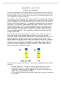
There are three key sites in a ribosome – aminoacyl (A), peptidyl (P) and exit (E).
Initiation:
Initiation factors (IF1, IF2 and IF3) and GTP bind to the small subunit.
The initiator tRNA with methionine binds to the P site, the subunit
recognises a binding sequence, upstream of the AUG start (methionine
codon). This causes the release of IF3.
GTP is hydrolysed allowing the binding of the large subunit, and release of
IF1 and IF2.
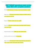
, Elongation:
Elongation factor-tu (EF-Tu) binds to charged tRNA molecules and attracts
the tRNA to the ribosome/mRNA complex.
When the appropriate charged tRNA (which has anticodon matching the
codon) binds to the codon in the A site, EF-Tu is released.
A peptide bond forms between the amino acids in the P and A sites. EF-G
binds and causes the ribosome to shift along the mRNA molecule, this
moves the position of the tRNA to the next site, causing the A site to be
free.
The tRNA in the E site dissociates, leaving a growing polypeptide chain,
attached to the complex by the peptide bond to the charged tRNA in the P
site.
Once the A site is free, the process continues in a cycle as the ribosome
moves along the mRNA. In bacteria about 20 amino acids can be added
every second.
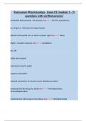
Termination:
When a stop codon is reached and present in the A site, instead of a tRNA
molecule being recruited, the codon is recognised by one of 3 release
factor proteins.
The binding of a release factor to the stop codon in the A site causes the
ribosome subunits and mRNA to dissociate, leaving all the components
free to start the process of translation again and a new polypeptide chain.
The triplet code allows the 4 possible bases in DNA (and RNA) to code for the 20
amino acids. There is redundancy in the code, an amino acid can have more than
one codon. This offers some resistance to some mutations having an effect, a
change in a base could still code for the same amino acid. This is known as a
silent point mutation. The triplet code does make a mutation where a nucleotide
is lost (deletion) or added (insertion) particularly disruptive. The loss or gain of
one nucleotide causes a frameshift mutation, which affects all the codons after
the insertion or deletion. These mutations usually have a big impact on the
protein to be expressed.
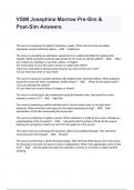
In bacteria, genes that code for related protein products are often grouped
together, in an operon. This allows proteins that have a related function to be
produced together. In the example below genes that code for enzymes involved
in the synthesis of tryptophan are together with a single promoter.
control of gene expression
The code in mRNA needs to be translated from the language of nucleic acids to
that of proteins. This occurs at the ribosomes. Ribosomes are large complexes of
protein and RNA. They are formed from two subunits, the small and large. They
act as platforms for protein synthesis.
The sequence of ribonucleotides that make up mRNA is read in groups of three, a
codon. By having the 4 possible nucleotides read in threes, there is the potential
to have 64 different codes, more than enough for 20 amino acids. This excess in
ability to code has an importance – the greater than required codes is known as
redundancy. The ribosome is the platform for protein synthesis. This occurs as
directed by the codons. The link between the instructions in the codon and the
amino acid to be made is by transfer RNA (tRNA). Enzymes called aminoacyl-
tRNA synthetases are responsible for catalysing the addition of amino acids to
the correct tRNA molecule. This is known as activation of the amino acid and
charging of the tRNA.
There are 61 coding codons (codons that code for amino acids) and there are
also 3 stop codons. Most species have the ability to produce 30-35 tRNA
molecules. There is some wobble allowed in the pairing between the codon and
anticodon pairing. Pairing in one position is not vital for all anticodon-codon
pairing. Because of the inclusion of inosine (a modified purine) in the 5’ position
of the anticodon on tRNA Ala1, it is capable of recognising 3 of the codons that
code for the amino acid alanine. tRNA Ala2 can only recognise one codon for
alanine. So, these two tRNA molecules can cover the 4 codons that code for
alanine, because of the wobble allowed.
There are three key sites in a ribosome – aminoacyl (A), peptidyl (P) and exit (E).
Initiation:
Initiation factors (IF1, IF2 and IF3) and GTP bind to the small subunit.
The initiator tRNA with methionine binds to the P site, the subunit
recognises a binding sequence, upstream of the AUG start (methionine
codon). This causes the release of IF3.
GTP is hydrolysed allowing the binding of the large subunit, and release of
IF1 and IF2.
, Elongation:
Elongation factor-tu (EF-Tu) binds to charged tRNA molecules and attracts
the tRNA to the ribosome/mRNA complex.
When the appropriate charged tRNA (which has anticodon matching the
codon) binds to the codon in the A site, EF-Tu is released.
A peptide bond forms between the amino acids in the P and A sites. EF-G
binds and causes the ribosome to shift along the mRNA molecule, this
moves the position of the tRNA to the next site, causing the A site to be
free.
The tRNA in the E site dissociates, leaving a growing polypeptide chain,
attached to the complex by the peptide bond to the charged tRNA in the P
site.
Once the A site is free, the process continues in a cycle as the ribosome
moves along the mRNA. In bacteria about 20 amino acids can be added
every second.
Termination:
When a stop codon is reached and present in the A site, instead of a tRNA
molecule being recruited, the codon is recognised by one of 3 release
factor proteins.
The binding of a release factor to the stop codon in the A site causes the
ribosome subunits and mRNA to dissociate, leaving all the components
free to start the process of translation again and a new polypeptide chain.
The triplet code allows the 4 possible bases in DNA (and RNA) to code for the 20
amino acids. There is redundancy in the code, an amino acid can have more than
one codon. This offers some resistance to some mutations having an effect, a
change in a base could still code for the same amino acid. This is known as a
silent point mutation. The triplet code does make a mutation where a nucleotide
is lost (deletion) or added (insertion) particularly disruptive. The loss or gain of
one nucleotide causes a frameshift mutation, which affects all the codons after
the insertion or deletion. These mutations usually have a big impact on the
protein to be expressed.
In bacteria, genes that code for related protein products are often grouped
together, in an operon. This allows proteins that have a related function to be
produced together. In the example below genes that code for enzymes involved
in the synthesis of tryptophan are together with a single promoter.