7.1 HAEMOGLOBIN
09 October 2020
09:54
Red blood cells contain haemoglobin.
The haemoglobins are a group of chemically similar molecules found in a wide
variety of organisms.
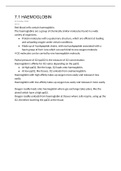
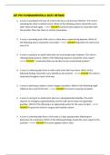
● Protein molecules with a quaternary structure, which are efficient at loading
and unloading oxygen under certain conditions.
● Made up of 4 polypeptide chains, with each polypeptide associated with a
haem group of Fe2+ ions which can each bind to one oxygen molecule.
4 O2 molecules can be carried by one haemoglobin molecule.
Partial pressure of O2 (ppO2) is the measure of O2 concentration.
Haemoglobin's affinity for O2 varies depending on the ppO2.
● At high ppO2, like the lungs, O2 loads onto haemoglobin.
● At low ppO2, like tissues, O2 unloads from oxyhaemoglobin.
Haemoglobin with high affinity takes up oxygen more easily and releases it less
easily.
Haemoglobin with low affinity takes up oxygen less easily and releases it more easily.
Oxygen readily loads onto haemoglobin where gas exchange takes place, like the
alveoli which have a high ppO2.
Oxygen readily unloads from haemoglobin at tissues where cells respire, using up the
O2, therefore lowering the ppO2 at the tissue.
, 7.2 TRANSPORT OF OXYGEN BY
HAEMOGLOBIN
13 March 2021
12:34
When haemoglobin is exposed to different ppO2, oxygen doesn't bind evenly.
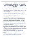
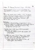
The graph of the relationship between haemoglobin saturation and ppO2 is known as
the O2 dissociation curve.
The reason for the curve being this shape is because:
● Shape of haemoglobin molecule makes it difficult for the first O2 to bind to
one of its 4 binding sites.
● When the first O2 molecule binds, it changes the quaternary structure and
therefore shape, inducing other subunits to bind to oxygen molecules.
● This positive cooperativity makes the binding of the 2nd and 3rd oxygen
molecules easier, so only a smaller increase in ppO2 is needed for the 2nd and
3rd oxygen to bind, causing the gradient to steepen.
● However, after the 3rd molecule binds, its harder for the 4th molecule to bind
due to probability. This is because it's less likely for the molecule to find the
empty binding site. The gradient reduces and the curve levels off.
The further left the curve, the greater the affinity of haemoglobin is for O2.
The further right the curve, the lower the affinity of haemoglobin for O2.
09 October 2020
09:54
Red blood cells contain haemoglobin.
The haemoglobins are a group of chemically similar molecules found in a wide
variety of organisms.
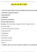
● Protein molecules with a quaternary structure, which are efficient at loading
and unloading oxygen under certain conditions.
● Made up of 4 polypeptide chains, with each polypeptide associated with a
haem group of Fe2+ ions which can each bind to one oxygen molecule.
4 O2 molecules can be carried by one haemoglobin molecule.
Partial pressure of O2 (ppO2) is the measure of O2 concentration.
Haemoglobin's affinity for O2 varies depending on the ppO2.
● At high ppO2, like the lungs, O2 loads onto haemoglobin.
● At low ppO2, like tissues, O2 unloads from oxyhaemoglobin.
Haemoglobin with high affinity takes up oxygen more easily and releases it less
easily.
Haemoglobin with low affinity takes up oxygen less easily and releases it more easily.
Oxygen readily loads onto haemoglobin where gas exchange takes place, like the
alveoli which have a high ppO2.
Oxygen readily unloads from haemoglobin at tissues where cells respire, using up the
O2, therefore lowering the ppO2 at the tissue.
, 7.2 TRANSPORT OF OXYGEN BY
HAEMOGLOBIN
13 March 2021
12:34
When haemoglobin is exposed to different ppO2, oxygen doesn't bind evenly.
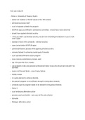
The graph of the relationship between haemoglobin saturation and ppO2 is known as
the O2 dissociation curve.
The reason for the curve being this shape is because:
● Shape of haemoglobin molecule makes it difficult for the first O2 to bind to
one of its 4 binding sites.
● When the first O2 molecule binds, it changes the quaternary structure and
therefore shape, inducing other subunits to bind to oxygen molecules.
● This positive cooperativity makes the binding of the 2nd and 3rd oxygen
molecules easier, so only a smaller increase in ppO2 is needed for the 2nd and
3rd oxygen to bind, causing the gradient to steepen.
● However, after the 3rd molecule binds, its harder for the 4th molecule to bind
due to probability. This is because it's less likely for the molecule to find the
empty binding site. The gradient reduces and the curve levels off.
The further left the curve, the greater the affinity of haemoglobin is for O2.
The further right the curve, the lower the affinity of haemoglobin for O2.