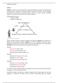
Microscopy
• Microscopes allow us to see things that we can’t see through our naked eye.
• There are two types of Microscopes: Electron & Light Microscope.
• Light microscope: uses light and lenses to focus on the image. Allows us to view cells &
larger components of the cell e.g. nucleus, cytoplasm e.tc. Light microscope was
invented in 1590s. They have low resolution as well as low magnification. The highest
magnification is x1500.
• Electron Microscope: Fires electrons at a sample to form an image. Magnification and
resolution are high.
• This means with an electron microscope you can zoom in way further and have a look at
things in more detail. You can see finer structures and sharp images.
• Electron microscopes are very expensive. We can see smaller things such as
mitochondria.
• However, electron microscopes do not detect color and they were invented in 1930s.
• There are two types of electron microscopes: Transmission & Scanning
• Transmission electron microscope: This produces the most magnified image. It fires the
beam of electrons at the sample. The beam allows the image to be produced by
focusing.
• Scanning Electron Microscope. It produces a 3D image of the sample. The beam of
electrons is fired at the sample with force that it goes across and is reflected to produce
the 3D image.
Light Microscope Electron Microscope
- Cheap - Expensive
- Small - Large
- Easy to carry - Hard to carry
- Simple - Complex
- Natural colored image - Black & white image
- Observes living or dead cells - Observes dead cells
- Resolution 0.2 m - Resolution 0.1 nanometer