Anatomie – Opmaat
ZSA VB Anatomie – lichaamswand
PowerPoint radiologie
Inleiding
- Voor tentamen: anatomische radiologie op beelden en kritieke bevindingen op een
thoraxfoto:
o Grote pneumothorax
o Grote atelectase
o Grote hoeveelheid pleuravocht
o Veel overvulling, decompensatio cordis
o Malpositie van de beademingstube
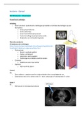
Normale anatomie
CT-abdomen in 3 richtingen
https://www.startpuntradiologie.nl/coschappen/spoedeisende-
hulp/buik/ct-abdomen-algemeen/index.html
- Transversaal
o Boven= voorkant patiënt
o Onder= achterkant
o Links foto= rechts patiënt
o Patiënt ligt en je bekijkt vanaf de voeten
- Coronaal
o Patiënt van voor naar achter
- Sagittaal
o Kijkt vanaf de zijkant
Casus 1
CT
- Door adipeus > organen goed te onderscheiden door tussenliggende vet
- Colontumor niet uit te sluiten met CT > dient coloscopie of voorbereide CT-colon-
Casus 2
CT
- Ribfracturen en hematopneumothorax
,Thoraxfoto
- Grote pneumothorax links
o Zie contour van gecollabeerde linkerlong
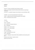
Casus 3
X-thorax
- Veel subcutaan emfyseem > hierdoor zijn
spiervezels van rechter m. pectoralis major af te
grenzen
- Geen pneumothorax R
- Thoraxdrain R
Casus 4
CTA na buikoperatie
- Er is in de ventrale buikwand een groot
hematoom zichtbaar onder de
operatienietjes.
- Zoek de a. mammaria links op: er is een
blush te zien.
o = bewijs voor bloeding
- Let op: ventrale buikwand
,CT veneuze fase
- Blush wordt groter > bewijs actieve bloeding
Angiografie
- De interventieradioloog heeft via de lies de katheter opgeschoven tot in de origo van de
linker a. mammaria (a. thoracica interna sinistra)
- Een microkatheter wordt opgeschoven via de katheter tot distaal in de a. mammaria/ a.
epigastrica superior tot vlak voor de blush.
- Via de microkatheter worden coils geplaatst waarna de bloeding is gestopt en de patiënt
weer hemodynamisch stabiel wordt
E-learning
CT
, MRA
ZSA VB Anatomie – lichaamswand
PowerPoint radiologie
Inleiding
- Voor tentamen: anatomische radiologie op beelden en kritieke bevindingen op een
thoraxfoto:
o Grote pneumothorax
o Grote atelectase
o Grote hoeveelheid pleuravocht
o Veel overvulling, decompensatio cordis
o Malpositie van de beademingstube
Normale anatomie
CT-abdomen in 3 richtingen
https://www.startpuntradiologie.nl/coschappen/spoedeisende-
hulp/buik/ct-abdomen-algemeen/index.html
- Transversaal
o Boven= voorkant patiënt
o Onder= achterkant
o Links foto= rechts patiënt
o Patiënt ligt en je bekijkt vanaf de voeten
- Coronaal
o Patiënt van voor naar achter
- Sagittaal
o Kijkt vanaf de zijkant
Casus 1
CT
- Door adipeus > organen goed te onderscheiden door tussenliggende vet
- Colontumor niet uit te sluiten met CT > dient coloscopie of voorbereide CT-colon-
Casus 2
CT
- Ribfracturen en hematopneumothorax
,Thoraxfoto
- Grote pneumothorax links
o Zie contour van gecollabeerde linkerlong
Casus 3
X-thorax
- Veel subcutaan emfyseem > hierdoor zijn
spiervezels van rechter m. pectoralis major af te
grenzen
- Geen pneumothorax R
- Thoraxdrain R
Casus 4
CTA na buikoperatie
- Er is in de ventrale buikwand een groot
hematoom zichtbaar onder de
operatienietjes.
- Zoek de a. mammaria links op: er is een
blush te zien.
o = bewijs voor bloeding
- Let op: ventrale buikwand
,CT veneuze fase
- Blush wordt groter > bewijs actieve bloeding
Angiografie
- De interventieradioloog heeft via de lies de katheter opgeschoven tot in de origo van de
linker a. mammaria (a. thoracica interna sinistra)
- Een microkatheter wordt opgeschoven via de katheter tot distaal in de a. mammaria/ a.
epigastrica superior tot vlak voor de blush.
- Via de microkatheter worden coils geplaatst waarna de bloeding is gestopt en de patiënt
weer hemodynamisch stabiel wordt
E-learning
CT
, MRA