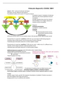
Molecular diagnostics COURSE 10BM
Lesson 1: MDX = advanced molecular techniques
Techniques used for diagnosis and prognosis
Translational medicine and research research proposal
Translation research: Translation of molecular
discoveries into clinical application (forward
translation).
Scientific questions that arise from relevant
clinical findings in turn triggers advances in
research (reverse translation)
Diagnosis:
Emergency presentation
• symptoms → poorer outcome
Screening
• Imaging:
Mammography (breast cancer)
Colonoscopy (colorectal cancer)
Laboratory tests
• PAP test (cervical cancer), CA-125 (ovarian cancer), PSA (prostate cancer), BRCA1 (breast cancer)
Characteristics of a good test: sensitivity (1/100 tests says not pregnant while being pregnant)
▪ Sensitive: detect small amounts, even in the presence of other molecules
▪ Sensitivity gives information about the % of false negative samples
Characteristics of a good test: specificity (1/100 tests say covid +, while in fact it’s a different virus)
▪ Specific: only the target molecule is detected (positive result)
▪ Specificity gives information about the % of false positive samples
Additional features of a good test: gives information about complex biology.
▪ High sensitivity and specificity: accuracy
▪ Potential for simple and standardized procedures
automation
▪ High throughput and cheap
<- The diversity of mutations that can initiate human cancer
Activating intragenic mutations: activating oncogenes
Inactivating intragenic mutations: gene is losing his functions (tumor suppressor genes) so cancer
Imaging:
Location, stage of tumor, growth, Plan treatment (localization of radiation) ▪ Monitor recurrence ▪
Intra vital imaging ▪ How drugs work and fail
Imaging systems vary in sensitivity and resolution
, Why are some patients resistant to therapy?
Why do some tumors recur more often?
Why and how do some tumors metastasize?
Clinical questions → translated to research
Why do some tumors grow faster than others? Proliferation rate may differ, angiogenesis, immune
respons, migration, apoptosis
Human epidermal growth factor receptor (HER2) on tissue and cellular level
HER2 gene amplification in 20-30% breast carcinomas
HER2 gene amplification → shorter survival / bad response to therapy
Need for treatment targeting HER2 receptor = Herceptin: monoclonal anti-HER2 antibody
Improved outcome for HER2 positive patients. Sometimes people don’t have a respons against
medicine, for example because of resistance
Imaging: drug discovery and functioning
Effect of drugs differ in vitro and in vivo
in vitro • 3D models • intra vital microscopy of live animals • Injectable imaging agents Quantification
of drug delivery and the effect of drug on single cells
Imaging in research: intra vital microscopy:
Drug target: tumor angiogenesis
• anti-VEGF antibodies
• Bevacizumab
Implantation of optical window Visualize if the drug inhibits angiogenesis
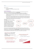
Imaging drug binding to target and problems thereof
Unravel why drugs fail: Imaging of drug binding (b in picture)
• Target tumor vasculature with peptide-functionalized vesicle
mAb directed against tumour cells cannot penetrate tumor limited uptake this appeared to
be the problem with HER2 therapy
The HER2 antibodies were labeled with a fluorescent probe, and it was visualized that the antibodies
do not penetrate the tumor mass fully (c in picture)
Drug does reach target Mixed expression of drug target (d in picture)
Drug targeting nucleus ends up in cytosol (e in picture)
Genetic reporter systems: FRET= Fluorescent resonance energy transfer
A method where distance dependent energy transfer is measured
Donor – acceptor (10-100A) = Overlapping excitation/emission spectra
a donor fluorophore absorbs the energy due to
excitation of incident light and transfers the excitation energy to a nearby
chromophore, the acceptor
What is measured? decreased of donor fluorescence and increase of acceptor
fluorescence
FRET can be used in chronic myeloid leukemia: translocation of BCR to ABL (fusion of chromosome 9
and 22) → fusion protein
▪ Higher and constitutively active tyrosine kinase, phosphorylation of CrKL associated with leukemia
▪ Treatment: tyrosine kinase inhibitor to stop the phosphorylation of CrKL
Fluorescent molecules are placed on both ends of CrKL, in normal situations no
phosphorylation (low fret), when it’s present phosphorylation will occur between BCR-
ABL and a conformational change happens (high fret). KNOW PRINCIPLE OF TECHNIQUE
Lesson 1: MDX = advanced molecular techniques
Techniques used for diagnosis and prognosis
Translational medicine and research research proposal
Translation research: Translation of molecular
discoveries into clinical application (forward
translation).
Scientific questions that arise from relevant
clinical findings in turn triggers advances in
research (reverse translation)
Diagnosis:
Emergency presentation
• symptoms → poorer outcome
Screening
• Imaging:
Mammography (breast cancer)
Colonoscopy (colorectal cancer)
Laboratory tests
• PAP test (cervical cancer), CA-125 (ovarian cancer), PSA (prostate cancer), BRCA1 (breast cancer)
Characteristics of a good test: sensitivity (1/100 tests says not pregnant while being pregnant)
▪ Sensitive: detect small amounts, even in the presence of other molecules
▪ Sensitivity gives information about the % of false negative samples
Characteristics of a good test: specificity (1/100 tests say covid +, while in fact it’s a different virus)
▪ Specific: only the target molecule is detected (positive result)
▪ Specificity gives information about the % of false positive samples
Additional features of a good test: gives information about complex biology.
▪ High sensitivity and specificity: accuracy
▪ Potential for simple and standardized procedures
automation
▪ High throughput and cheap
<- The diversity of mutations that can initiate human cancer
Activating intragenic mutations: activating oncogenes
Inactivating intragenic mutations: gene is losing his functions (tumor suppressor genes) so cancer
Imaging:
Location, stage of tumor, growth, Plan treatment (localization of radiation) ▪ Monitor recurrence ▪
Intra vital imaging ▪ How drugs work and fail
Imaging systems vary in sensitivity and resolution
, Why are some patients resistant to therapy?
Why do some tumors recur more often?
Why and how do some tumors metastasize?
Clinical questions → translated to research
Why do some tumors grow faster than others? Proliferation rate may differ, angiogenesis, immune
respons, migration, apoptosis
Human epidermal growth factor receptor (HER2) on tissue and cellular level
HER2 gene amplification in 20-30% breast carcinomas
HER2 gene amplification → shorter survival / bad response to therapy
Need for treatment targeting HER2 receptor = Herceptin: monoclonal anti-HER2 antibody
Improved outcome for HER2 positive patients. Sometimes people don’t have a respons against
medicine, for example because of resistance
Imaging: drug discovery and functioning
Effect of drugs differ in vitro and in vivo
in vitro • 3D models • intra vital microscopy of live animals • Injectable imaging agents Quantification
of drug delivery and the effect of drug on single cells
Imaging in research: intra vital microscopy:
Drug target: tumor angiogenesis
• anti-VEGF antibodies
• Bevacizumab
Implantation of optical window Visualize if the drug inhibits angiogenesis
Imaging drug binding to target and problems thereof
Unravel why drugs fail: Imaging of drug binding (b in picture)
• Target tumor vasculature with peptide-functionalized vesicle
mAb directed against tumour cells cannot penetrate tumor limited uptake this appeared to
be the problem with HER2 therapy
The HER2 antibodies were labeled with a fluorescent probe, and it was visualized that the antibodies
do not penetrate the tumor mass fully (c in picture)
Drug does reach target Mixed expression of drug target (d in picture)
Drug targeting nucleus ends up in cytosol (e in picture)
Genetic reporter systems: FRET= Fluorescent resonance energy transfer
A method where distance dependent energy transfer is measured
Donor – acceptor (10-100A) = Overlapping excitation/emission spectra
a donor fluorophore absorbs the energy due to
excitation of incident light and transfers the excitation energy to a nearby
chromophore, the acceptor
What is measured? decreased of donor fluorescence and increase of acceptor
fluorescence
FRET can be used in chronic myeloid leukemia: translocation of BCR to ABL (fusion of chromosome 9
and 22) → fusion protein
▪ Higher and constitutively active tyrosine kinase, phosphorylation of CrKL associated with leukemia
▪ Treatment: tyrosine kinase inhibitor to stop the phosphorylation of CrKL
Fluorescent molecules are placed on both ends of CrKL, in normal situations no
phosphorylation (low fret), when it’s present phosphorylation will occur between BCR-
ABL and a conformational change happens (high fret). KNOW PRINCIPLE OF TECHNIQUE