Nutrition and energy – Peptic ulcer disease (PUD) #1
Clinical definitions Sites of ulcer formation Pathophysiology
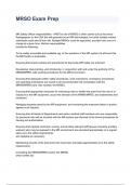
Peptic ulcer disease (PUD) refers to an ulcer in the
lower oesophagus, stomach or duodenum. It the
jejunum, ulcers can occur after surgical
anastomosis.
Ulceration vs Erosion
Erosion Ulceration
Most common site – Superior duodenum
Penetrates the Penetrates the
mucosa but not the mucosa and the Least common site – Oesophagus
By far the most common cause of
muscularis mucosa muscularis mucosa
PUD is helicobacter Pylori bacterial
Clinical presentation infection
Other risk factors include:
Recurrent episodes of abdominal pain NSAIDS - long term use damages
localised to the epigastrium. gastric mucosal defences
• This pain occurs in patterns with food. Smoking – impairs sufficient healing
of formed ulcers.
• Patients can present with vomiting
• Patients can also present with anorexia
and nausea, or early satiety after meals
• In some patients, the ulcer can be
‘silent’, presenting for the first time with
anaemia from chronic blood loss
• Anaemia – From pernicious anaemia
, Nutrition and energy – Peptic ulcer disease (PUD) #2
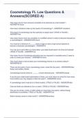
Helicobacter pylori Pathophysiology (continued) Normal function of gastric
mucosal cells
Very prominent in eastern Europe and Asia
Parietal cells secrete HCL and intrinsic factor
Chief cells secrete pepsinogen and gastric
lipase
G cells are a type of enteroendocrine cell which
secrete gastrin
Stimulants of gastric acid secretion
Greater prominence in eastern Europe and Aisa
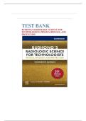
Investigations Management
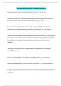
Virulence factors of H.pylori:
Flagella – giving the bacterium motility Most common investigation:
to burrow to the surface epithelial layer, The H.Pylori stool antigen test Triple therapy
beneath the mucus barrier. eradication
Other tests:
Adhesins – proteins which allow the Urease breath test – give pt 2 of 3 antibiotics for 1
radiolabelled urea to drink, week: H. pylori colonization in pyloric antrum:
bacteria to bind to antigens on the
which produces radiolabelled Initial gastrin secretion subsequent HCl secretion
surface of gastric epithelial cells (where
CO2 Clarithromycin (Antral gastritis – duodenal ulcer formation)
the PH is almost neutral)
Metronidazole
Biopsy and CLO petri dish test Amoxycillin Followed by: Chronic atrophic gastritis (whole
Urease – An enzyme secreted by this
(sample placed in dish with stomach): Inflammation and scarring leads to
bacterium to convert urea to ammonia.
urea, PH indicator turns from + One PPI for 1 week: destruction of parietal cells and hypochlorhydria
This raises the PH surrounding the
bacteria, buffering the harmful yellow to red) Lansoprazole
acidic environment. Omeprazole
Clinical definitions Sites of ulcer formation Pathophysiology
Peptic ulcer disease (PUD) refers to an ulcer in the
lower oesophagus, stomach or duodenum. It the
jejunum, ulcers can occur after surgical
anastomosis.
Ulceration vs Erosion
Erosion Ulceration
Most common site – Superior duodenum
Penetrates the Penetrates the
mucosa but not the mucosa and the Least common site – Oesophagus
By far the most common cause of
muscularis mucosa muscularis mucosa
PUD is helicobacter Pylori bacterial
Clinical presentation infection
Other risk factors include:
Recurrent episodes of abdominal pain NSAIDS - long term use damages
localised to the epigastrium. gastric mucosal defences
• This pain occurs in patterns with food. Smoking – impairs sufficient healing
of formed ulcers.
• Patients can present with vomiting
• Patients can also present with anorexia
and nausea, or early satiety after meals
• In some patients, the ulcer can be
‘silent’, presenting for the first time with
anaemia from chronic blood loss
• Anaemia – From pernicious anaemia
, Nutrition and energy – Peptic ulcer disease (PUD) #2
Helicobacter pylori Pathophysiology (continued) Normal function of gastric
mucosal cells
Very prominent in eastern Europe and Asia
Parietal cells secrete HCL and intrinsic factor
Chief cells secrete pepsinogen and gastric
lipase
G cells are a type of enteroendocrine cell which
secrete gastrin
Stimulants of gastric acid secretion
Greater prominence in eastern Europe and Aisa
Investigations Management
Virulence factors of H.pylori:
Flagella – giving the bacterium motility Most common investigation:
to burrow to the surface epithelial layer, The H.Pylori stool antigen test Triple therapy
beneath the mucus barrier. eradication
Other tests:
Adhesins – proteins which allow the Urease breath test – give pt 2 of 3 antibiotics for 1
radiolabelled urea to drink, week: H. pylori colonization in pyloric antrum:
bacteria to bind to antigens on the
which produces radiolabelled Initial gastrin secretion subsequent HCl secretion
surface of gastric epithelial cells (where
CO2 Clarithromycin (Antral gastritis – duodenal ulcer formation)
the PH is almost neutral)
Metronidazole
Biopsy and CLO petri dish test Amoxycillin Followed by: Chronic atrophic gastritis (whole
Urease – An enzyme secreted by this
(sample placed in dish with stomach): Inflammation and scarring leads to
bacterium to convert urea to ammonia.
urea, PH indicator turns from + One PPI for 1 week: destruction of parietal cells and hypochlorhydria
This raises the PH surrounding the
bacteria, buffering the harmful yellow to red) Lansoprazole
acidic environment. Omeprazole