Osmoregulation (control of blood water
potential)
It is vital for all animals to control the water potential (Ψ) of their blood. This is because blood
with a low water potential will cause water to leave surrounding cells and tissues and enter the
blood by osmosis, whereas blood with high Ψ will cause water to leave the blood and enter the
tissues by osmosis. This causes shrinking or bursting of surrounding cells.
Water potential of the blood is affected by concentrations of solutes (ions, glucose, etc),
respiratory rate, amount of water consumed, etc.
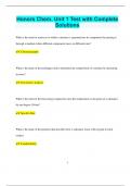
Structure of the kidney
The kidney has several main
sections, each with a different
function.
Fibrous capsule = outer membrane
that protects the kidney.
Cortex = outer region of the kidney
containing renal capsules, PCT,
DCT and blood vessels.
Medulla = inner region containing
loops of Henle, collecting ducts and
blood vessels.
Renal pelvis = a ‘funnel’ that
collects and directs urine into the
ureter.
Ureter = carries urine to the
bladder.
Renal artery = supplies the kidney with blood from the aorta. Linked to the afferent arteriole.
Renal vein = returns blood to the heart via the vena cava. Linked to the efferent arteriole.
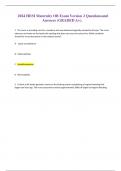
, Structure of the nephron
Nephrons are the functional
units of the kidney. Each
section of a nephron has
adaptations for their specific
function.
Renal capsule = a
cup-shaped structure
containing the glomerulus
(a knot of capillaries) with
podocytes lining its inner
layer.
Proximal convoluted tubule
(PCT) = a tube forming a
series of loops surrounded
by capillaries. Its walls have
microvilli.
Loop of Henle = a hairpin
loop surrounded by
capillaries.
Distal convoluted tubule (DCT) = a series of loops surrounded by fewer blood capillaries than
the PCT.
Collecting duct = a tube into which many DCTs empty their filtrate. It ends in the renal pelvis.
Afferent arteriole = a vessel supplying the nephron with blood from the renal artery.
Efferent arteriole = a vessel which collects blood from the glomerulus and becomes many
capillaries, eventually leading into the renal vein. Smaller diameter than the afferent arteriole.
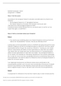
The osmoregulation process
Forming the glomerular filtrate via ultrafiltration
Blood enters the kidney via the renal artery which forms the afferent arterioles. Each of these
afferent arterioles enters a renal capsule, where it divides into the glomerulus. This is where
ultrafiltration occurs.
1. CREATING HYDROSTATIC PRESSURE
The afferent arteriole has a wider diameter than the efferent arteriole, so the hydrostatic
pressure in the glomerular capillaries is increased.
2. ULTRAFILTRATION
potential)
It is vital for all animals to control the water potential (Ψ) of their blood. This is because blood
with a low water potential will cause water to leave surrounding cells and tissues and enter the
blood by osmosis, whereas blood with high Ψ will cause water to leave the blood and enter the
tissues by osmosis. This causes shrinking or bursting of surrounding cells.
Water potential of the blood is affected by concentrations of solutes (ions, glucose, etc),
respiratory rate, amount of water consumed, etc.
Structure of the kidney
The kidney has several main
sections, each with a different
function.
Fibrous capsule = outer membrane
that protects the kidney.
Cortex = outer region of the kidney
containing renal capsules, PCT,
DCT and blood vessels.
Medulla = inner region containing
loops of Henle, collecting ducts and
blood vessels.
Renal pelvis = a ‘funnel’ that
collects and directs urine into the
ureter.
Ureter = carries urine to the
bladder.
Renal artery = supplies the kidney with blood from the aorta. Linked to the afferent arteriole.
Renal vein = returns blood to the heart via the vena cava. Linked to the efferent arteriole.
, Structure of the nephron
Nephrons are the functional
units of the kidney. Each
section of a nephron has
adaptations for their specific
function.
Renal capsule = a
cup-shaped structure
containing the glomerulus
(a knot of capillaries) with
podocytes lining its inner
layer.
Proximal convoluted tubule
(PCT) = a tube forming a
series of loops surrounded
by capillaries. Its walls have
microvilli.
Loop of Henle = a hairpin
loop surrounded by
capillaries.
Distal convoluted tubule (DCT) = a series of loops surrounded by fewer blood capillaries than
the PCT.
Collecting duct = a tube into which many DCTs empty their filtrate. It ends in the renal pelvis.
Afferent arteriole = a vessel supplying the nephron with blood from the renal artery.
Efferent arteriole = a vessel which collects blood from the glomerulus and becomes many
capillaries, eventually leading into the renal vein. Smaller diameter than the afferent arteriole.
The osmoregulation process
Forming the glomerular filtrate via ultrafiltration
Blood enters the kidney via the renal artery which forms the afferent arterioles. Each of these
afferent arterioles enters a renal capsule, where it divides into the glomerulus. This is where
ultrafiltration occurs.
1. CREATING HYDROSTATIC PRESSURE
The afferent arteriole has a wider diameter than the efferent arteriole, so the hydrostatic
pressure in the glomerular capillaries is increased.
2. ULTRAFILTRATION