100% satisfaction guarantee Immediately available after payment Both online and in PDF No strings attached 4.2 TrustPilot
logo-home
Exam (elaborations)

BIOD 151 / BIOD151 ANATOMY AND PHYSIOLOGY MODULE 5

Rating
-
Sold
-
Pages
149
Grade
A+
Uploaded on
04-05-2022
Written in
2021/2022

BIOD 151 / BIOD151 ANATOMY AND PHYSIOLOGY MODULE 5 Anatomy of the Muscular System: Introduction & Muscles of the Head, Neck, and Trunk Notice: To optimize your learning in this course, we advise that you complete the labs and modules as indicated in the BIOD 151 Lab Schedule. The muscular system and the skeletal system work together to provide movement for the human body. Muscle tissue is found in three distinct types in the body; skeletal, smooth, and cardiac. Skeletal muscles must have a blood and nervous supply to provide movement. Skeletal muscles are under conscious control, meaning that a person can consciously decide to use these muscles to complete an action. Communication within the body to coordinate movement starts in the brain with a message that is sent through the spinal cord and eventually attaches to a muscle. Peripheral nerves carry the signal from the central nervous system (brain and spinal cord) to a specific muscle destination to provide movement. Messages from the central nervous system to a muscle are called a motor actions. Nerves also carry information from the external environment to the central nervous system, called sensation or sensory input. (see Figure 5.1 and Figure 5.2) Spinal nerves combine to form complex networks of peripheral nerves throughout the body. Figure 5.1 Posterior view of the nervous system. The brain and spinal cord (central nervous system) connect to the peripheral nervous system. Examples of peripheral nerves are spinal nerves (cervical, thoracic, and lumbar), the upper trunk of the brachial plexus, and the radial nerve. Figure 5.2 Peripheral nerves carry the communication from the central nervous system (brain and spinal cord) to the muscle. Peripheral nerves also carry information from the environment to the central nervous system. Tendons are connective tissues that connect skeletal muscle to bone at each end (see Figure 5.3). Tendons are flexible, can bend at the joints, and help cushion against sudden movement. Ligaments are connective tissue that connects bone to bone, helping to stabilize joints where bones meet. Figure 5.3 Skeletal muscles attach to bones via tendons at points called the origin and insertion. The origin is the fixed point while the insertion is the place that is moved during a muscle contraction. All skeletal muscles have an origin, insertion, and action. The origin is the bony site of attachment which is stationary during the movement. The insertion of a muscle is the bony site of attachment that is moved by the muscle contraction. (see Figure 5.3) The origin and insertion can be reversed in different types of movement. For this module, the standard origin and insertion points are referenced from anatomical position. The action of the muscle is what effect is produced by the muscle’s contraction. For example, the triceps is the primary extensor of the forearm. The innervation is the peripheral nerve that supplies a muscle with the message from the brain. For example, the innervation of the biceps brachii is the musculocutaneous nerve. (see Figure 5.4) Figure 5.4 Anterior view of the musculocutaneous nerve (blue) innervates the biceps brachii muscle. Muscles are generally studied in groups. The following muscle list is an overview of some of the major muscles in the body. Study the location of the muscle in the body. In addition, if listed, study the origin, insertion, innervation and action of these muscles. Muscles of Facial Expression: (See Figure 5.5) The muscles in this grouping help to facilitate all the actions within the face. All the varied facial expressions are enabled by these muscles. CN is an abbreviation for cranial nerve. There are twelve pairs of cranial nerves that originate on the ventral surface of the brain, controlling muscles and functions of several organs and glands. Orbicularis oculi Action: eye closure Innervation: Facial nerve (CN VII) Orbicularis oris Action: mouth closure: closes lips, protrudes lips forward, presses lips against teeth Innervation: Facial nerve (CN VII) Zygomaticus major/minor Action: pull corners of lips upward Innervation: Facial nerve (CN VII) Risorius Action: pulls the corners of the mouth posteriorly (grin or grimace) Innervation: Facial nerve (CN VII) Frontalis (occipitofrontailis) Action: raise eyebrows Innervation: Facial nerve (CN VII) Buccinator Action: compress cheeks Innervation: Facial nerve (CN VII) Masseter Action: jaw closure Innervation: Trigeminal nerve (CN V, mandibular branch) Temporalis Action: Elevates mandible, closes jaw Innervation: Trigeminal nerve (CN V, mandibular branch) Figure 5.5 Muscles of facial expression. Note that the buccinator is deep to zygomaticus major/minor and risorius. Muscles of the Head and Neck: (See Figure 5.6) The muscles in this grouping help to facilitate actions which move the head (occiput) and control the neck. Side bending of the neck is also called side flexion or lateral flexion, which refers to moving one of the ears to the same side shoulder. Neck flexion refers to the action of moving the chin towards the sternum. Note there are several muscles that have differing actions if acting together on both sides (bilaterally) or acting on one side only (unilaterally). Semispinalis Capitis Origin: Articular processes of inferior cervical & transverse process of superior thoracic vertebrae Insertion: Occipital bone Action: Bilateral extend head Unilateral laterally flexes neck to same side Innervation: Spinal nerves Sternocleidomastoid Origin: Sternal end of clavicle and manubrium Insertion: Mastoid region of skull Action: Bilateral: Neck flexion Unilateral: Turns face to opposite side Innervation: Accessory nerve (CN XI) Splenius Capitis Origin: Spinous process/ligaments of inferior cervical vertebrae Insertion: Mastoid process, occipital bones of skull Action: Bilateral extend head Unilateral laterally flexes neck to same side Innervation: Cervical spinal nerves Longissimus cervicis Origin: transverse processes of superior thoracic vertebrae Insertion: transverse process of middle and superior cervical vertebrae Action: Bilateral extend head Unilateral laterally flexes neck to same side Innervation: Cervical and thoracic spinal nerves Longissimus thoracis Origin: Transverse process of inferior thoracic, superior lumbar vertebrae Insertion: Transverse process of middle and superior cervical vertebrae Action: Bilateral extend head Unilateral laterally flexes neck to same side Innervation: Thoracic and lumbar spinal nerves Thyrohyoid Origin: Thyroid cartilage of larynx Insertion: Hyoid bone Action: Elevates thyroid, depresses hyoid bone Innervation: Hypoglossal nerve Scalenes (anterior, middle, posterior) Origin: Transverse processes of C2- C7 Insertion: 1st and 2nd Ribs Action: Elevates ribs 1 & 2 Innervation: Cervical spinal nerves Figure 5.6 Lateral view: Muscles of the head and neck Vertebral Column: Erector Spinae (See Figure 5.7) The erector spinae group are superficial muscles in the back. These muscles act as the prime extensors of the vertebral column. Flexion of the spine refers to forward bending the trunk (such as a “cat” stretch). Extension of the spine refers backward bending. The vertebral column is also able to perform side bending (or lateral flexion) as well as movement around an axis (rotation). Spinalis: Medial column of erector spinae Made up of three divisions (spinalis thoracis, spinalis cervicis, and spinalis capitis) Longissimus: Intermediate column of erector spinae Made up of three divisions (longissimus dorsi, longissimus cervicis, longissimus capitits) Iliocostalis: Lateral column of erector spinae Made up of three divisions (iliocostalis lumborum, iliocostalis thoracis, iliocostalis cervicis) Figure 5.7 Posterior view: Erector Spinae Group Oblique and rectus muscles: Anterior (See Figure 5.8) The abdominal muscles perform many functions. These muscles help to support and protect the abdominal viscera (which are not protected by bony structures). These muscles, along with the muscles in the back (see above) help to provide support for upright posture and balance. Rectus abdominis Origin: Pubic Crest, pubic symphysis Insertion: Pubis, cartilages of ribs 5-7, xiphoid process Action: Flexion of spine, compression of abdominal viscera Innervation: Spinal nerves (T 7-T 12) External oblique Origin: Sternum, ribs (5-12) Insertion: Linea alba, iliac crest Action: Bilaterally: Flexion of the spine, compress the abdomen; Unilaterally: Flexes trunk to same side, rotates to opposite side Innervation: Lower intercostal, ilioinguinal nerves Internal oblique Origin: Iliac crest, lumbodorsal fascia Insertion: Inferior ribs, linea alba Action: Compresses abdomen, depresses ribs, flexes spine Innervation: Lower intercostal, ilioinguinal nerves Transverse abdominis – deep to internal oblique Origin: Lateral inguinal ligament, inner iliac crest Insertion: Linea alba, pubis Action: Compression of abdomen Innervation: First lumbar nerve (T 7- L1), iliohypogastric (T12-L1), ilioinguinal (T12-L1) Figure 5.8 Muscles and landmarks of the anterior trunk Muscles of Breathing (See Figure 5.9) The diaphragm is the primary muscle necessary for intaking air. The internal intercostals and the external intercostals work in opposite functions. Notice the difference in their muscle fiber orientation (see Figure 5.9). Diaphragm Origin: Cartilage of ribs 7-12, xiphoid process, lumbar vertebrae Insertion: Anterior longitudinal ligament (vertebral column) Action: Expands thoracic cavity, compresses abdominal cavity Innervation: Phrenic nerve (C3-5) Internal Intercostals Origin: Superior border of ribs 2-12 Insertion: Inferior of ribs above (1-11) Action: Depresses ribs (forced expiration) Innervation: Intercostal nerves External Intercostals Origin: Lower border of ribs 1-11 Insertion: Upper border of ribs below (1-12) Action: Elevates ribs (normal inspiration) Innervation: Intercostal nerves Figure 5.9 Anterior view of the muscles of breathing. The diaphragm is the primary muscle of inspiration. Anatomy of the Muscular System Part II: Muscles of the Upper and Lower Extremities Muscles of the Shoulder and Upper Extremity (See Figures 5.10, 5.11, 5.12) The muscles on the posterior thorax assist in movement of the shoulder girdle. The trapezius is a large muscle that acts on the shoulder as well as the neck and upper spine. The trapezius is superficial to the remaining muscles shown (see Figure 5.10). Trapezius (upper, middle, lower) Origin: Occipital bone, spinous process of T1-12, Insertion: Lateral clavicle, acromion, and scapular spine of scapula Action: Rotation, retraction, elevation, depression of scapula; extends neck; stabilizes shoulder Innervation: Accessory nerve (Cranial Nerve 11) Levator scapulae Origin: Transverse process of C1-4 Insertion: Medial border of scapula Action: Elevates scapula Innervation: Dorsal scapular nerve Rhomboids (major, minor) Origin: Spinous process (minor: C7-T1) (major: T2-5) Insertion: Medial border of scapula Action: Retraction of scapula Innervation: Dorsal scapular nerve Figure 5.10 Posterior view of the shoulder girdle muscles. The trapezius is a superficial muscle (shown on left). On the right, the trapezius is cut away to show the muscles underneath. *Note: Additional muscles are labeled in this figure for clarification. Pectoralis minor Origin: Ribs 3-5 Insertion: Coracoid process of scapula Action: Elevates ribs, draws scapula down and medially Innervation: Medial pectoral nerve Serratus Anterior Origin: Upper 8-9 ribs Insertion: Medial border of scapula Action: Protraction of scapula Innervation: Long thoracic nerve Figure 5.11 Anterior view of the muscles of the thorax and shoulder girdle. Serratus anterior (left and right) is highlighted in blue. Muscles of the shoulder girdle continued: (See Figures 5.12, 5.13, 5.14) The shoulder is a ball and socket joint, which permits many types of movement. The glenoid cavity of the scapula is very small compared to the head of the humerus. The large bony structure of the shoulder requires many ligaments and layers of muscle for stability. A muscle grouping known as the rotator cuff is necessary to stabilize the humeral head inside the shoulder joint. The rotator cuff is made up of the “SITS” muscles: Supraspinatus, Infraspinatus, Teres minor, and Subscapularis. (See Figure 5.14) Deltoid (anterior, posterior, middle) Origin: Clavicle and scapula Insertion: Deltoid tuberosity of humerus Action: Abduction at shoulder (whole muscle) Innervation: Axillary nerve Pectoralis major Origin: Ribs 2-6, body of sternum Insertion: Greater tubercle of humerus Action: Flexion, adduction and medial rotation at shoulder Innervation: Pectoral Nerves Supraspinatus- (above spine of scapula) Origin: Supraspinatus fossa of scapula Insertion: Greater tubercle of humerus Action: Abduction at the shoulder Innervation: Suprascapular nerve Infraspinatus (below spine of scapula) Origin: Infraspinatus fossa of scapula Insertion: Greater tubercle of humerus Action: Lateral rotation at shoulder Innervation: Suprascapular nerve Teres Minor Origin: Lateral border of scapula Insertion: Greater tubercle of humerus Action: Lateral rotation at shoulder Innervation: Axillary nerve Teres Major – not part of the rotator cuff Origin: Inferior angle of scapula Insertion: Intertubercular groove of humerus Action: Extension, adduction, and medial rotation at shoulder Innervation: Lower subscapular nerve Subscapularis- along inner surface of the scapula Origin: Subscapular fossa of scapula Insertion: Lesser tubercle of humerus Action: Medial rotation at the shoulder Innervation: Subscapular nerves Latissimus Dorsi Origin: Spinous process of inferior thoracic and lumbar vertebrae, ribs 8-12 Insertion: Intertubercular groove of humerus Action: Extension, adduction, and medial rotation at shoulder Innervation: Thoracodorsal nerve Coracobrachialis Origin: Coracoid process of scapula Insertion: Medial shaft of humerus Action: Adduction and flexion at shoulder Innervation: Musculocutaneous nerve

Show more Read less
Institution
Course


Whoops! We can’t load your doc right now. Try again or contact support.

Written for

Institution
Course

Document information

Uploaded on
May 4, 2022
Number of pages
149
Written in
2021/2022
Type
Exam (elaborations)
Contains
Questions & answers

Subjects

Content preview

BIOD 151 / BIOD151 ANATOMY AND PHYSIOLOGY
MODULE 5
Anatomy of the Muscular System: Introduction & Muscles of the Head,
Neck, and Trunk

Notice: To optimize your learning in this course, we advise that you complete the labs
and modules as indicated in the BIOD 151 Lab Schedule.


The muscular system and the skeletal system work together to provide movement for
the human body. Muscle tissue is found in three distinct types in the body; skeletal,
smooth, and cardiac.
Skeletal muscles must have a blood and nervous supply to provide movement. Skeletal
muscles are under conscious control, meaning that a person can consciously decide to
use these muscles to complete an action. Communication within the body to coordinate
movement starts in the brain with a message that is sent through the spinal cord and
eventually attaches to a muscle. Peripheral nerves carry the signal from the central
nervous system (brain and spinal cord) to a specific muscle destination to provide
movement. Messages from the central nervous system to a muscle are called a motor
actions. Nerves also carry information from the external environment to the central
nervous system, called sensation or sensory input. (see Figure 5.1 and Figure 5.2) Spinal
nerves combine to form complex networks of peripheral nerves throughout the body.

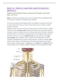
,Figure 5.1 Posterior view of the nervous system. The brain and spinal cord (central nervous

,system) connect to the peripheral nervous system. Examples of peripheral nerves are spinal nerves
(cervical, thoracic, and lumbar), the upper trunk of the brachial plexus, and the radial nerve.


Figure 5.2 Peripheral nerves carry the communication from the central nervous system (brain and
spinal cord) to the muscle. Peripheral nerves also carry information from the environment to the
central nervous system.


Tendons are connective tissues that connect skeletal muscle to bone at each end (see Figure 5.3).
Tendons are flexible, can bend at the joints, and help cushion against sudden
movement. Ligaments are connective tissue that connects bone to bone, helping to
stabilize joints where bones meet.

, Figure 5.3 Skeletal muscles attach to bones via tendons at points called the origin and insertion.
The origin is the fixed point while the insertion is the place that is moved during a muscle
contraction.


All skeletal muscles have an origin, insertion, and action. The origin is the bony site of
attachment which is stationary during the movement. The insertion of a muscle is the
bony site of attachment that is moved by the muscle contraction. (see Figure 5.3)


The origin and insertion can be reversed in different types of movement. For this
module, the standard origin and insertion points are referenced from anatomical
position.


The action of the muscle is what effect is produced by the muscle’s contraction. For
example, the triceps is the primary extensor of the forearm. The innervation is the
peripheral nerve that supplies a muscle with the message from the brain. For example,
the innervation of the biceps brachii is the musculocutaneous nerve. (see Figure 5.4)

Get to know the seller

Seller avatar
Reputation scores are based on the amount of documents a seller has sold for a fee and the reviews they have received for those documents. There are three levels: Bronze, Silver and Gold. The better the reputation, the more your can rely on the quality of the sellers work.
Marilyn77 kinuwrites
Follow You need to be logged in order to follow users or courses
Sold
170
Member since
4 year
Number of followers
140
Documents
334
Last sold
1 month ago
EXAMS DIRECTORY

Are you having problems with your exams? There are better ways to cope with that. Exams, notes, case studies, Testbanks, and many more are available here. The ability to not know something is the knowledge to know something better. Good luck with your studies. Hit me up if you need anything else Assignments, Case Studies, Research, Essay writing service, Questions and Answers, Discussions, etc. FOR THE BEST EXAMS AND VERIFIED ANSWERS GUARANTEED A+

Read more Read less
4.3

31 reviews

5
17
4
9
3
4
2
0
1
1

Recently viewed by you

Why students choose Stuvia

Created by fellow students, verified by reviews

Quality you can trust: written by students who passed their tests and reviewed by others who've used these notes.

Didn't get what you expected? Choose another document

No worries! You can instantly pick a different document that better fits what you're looking for.

Pay as you like, start learning right away

No subscription, no commitments. Pay the way you're used to via credit card and download your PDF document instantly.

Student with book image

“Bought, downloaded, and aced it. It really can be that simple.”

Alisha Student

Frequently asked questions