Physiology lecture P1
Eicosanoids synthesis: a role in autocrine signalling, involved in pain, fever and inflammation.
Transmembrane proteins:
- Ion-channel-linked receptor
- G-protein-linked receptor
- Enzyme-linked receptor
Synaptic signalling
Trimeric GTP-binding
Signalling by phosphorylation & signalling by GTP-binding protein
APP = ADP, GPP = GDP
Signal integration, converging of pathways
Downstream signals
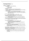
Gs couples receptor activation to cAMP formation
cAMP activates A-kinase, which activates phosphorylase kinase, which activates glycogen
phosphorylase, resulting in glycogen conversion to glucose-I-phosphate, i.e. glycolysis.
Calcium homeostasis
Calcium-binding molecules
25 receptor mediated processes via G-protein-linked receptor and phospholipase-C-
(kringel s), which cleaves PIP2.
C-kinase activation results in gene transcription.
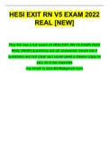
Enzyme-linked cell surface receptors: most receptor for growth and differentiation factors
are transmembrane tyrosine-specific protein kinases.
MAP-kinase(-kinase) (-kinase)
Tyrosine phosphorylation and subsequent SH2 interaction with messenger protein leads to
activation of Ras proteins, all of these reactions are quickly reversed.
Long-lived relays to the nucleus involve multiple cascades of serine/ theonine
phophorylations, esp. performed by the MAP (mitogen-activated protein) kinases (or ERK -
extracellular-signal-regulated kinases)
Epithelium(cell) including glands
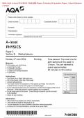
Cell adhesion molecules (CAM’s)
1. Cadherins: cell-cell adhesion
2. Selectins: temporary cell-cell connections
3. Integrins: cell-matrix adhesion
4. Immunoglobulin (Ig) – like CAM’s: cell-cell connections
Cadherins form a kind of zipper to bind a cell to a cell, you have different types, they can
steer the faith of cells by themselves.
Selectins are important in adhesion of leukocytes during inflammatory processes.
, Endothelium lines our blood vessels.
Weak adhesion and rolling (selectin-dependent) & strong adhesion and emigration (integrin-
dependent).
Adhesion to the extracellular matrix is achieved through integrins (focal adhesions).
IG-like adhesion molecules also can connect cells to each other.
Epithelium forms an interconnected layer due to cell-cell connections and the cell-matrix
connections.
Gap junctions allow the passage of small water-soluble molecules from cell to cell.
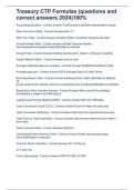
Cell connecting structures
1. Adhesion structures: attachment between cells or between cells and matrix
- Intermediate filaments, desmosomes or macula adherens hemidesmosomes
- Microfilaments actin filaments, zonula adherens (band shaped), adhesion plates (also
called focal adhesions)
- Adhesion of cells to cells or matrix are mediated by anchor proteins which serve to
couple the CAM’s to intracellular filaments.
- Zonula adherens uses cadherins to connect the cells and connects to actin filaments.
2. Occludens connections: closing connections, seal of the cell layer -> tight junction
(zonula occludens) (closes the space between two cells)
- passage of molecules occurs through the cytoplasm of the epithelial cell.
3. Communication connections: communication among cells
- Synapses (muscle – nerve cell, nerve cells)
- Nexus connections (gap junctions)
Lamina basalis and basal membrane
Lamina basalis
- Extracellular, presents the border between connective tissue and other tissue types.
- Function is adhesion and sieve/ filter
- EM: lamina densa (dark, collagen type 4 and proteoglycans), flanked by lamina lucida
on both sides (brighter, laminin)
- Lamina basalis consists of type 4 collagen, with glycoproteins such as laminin to
which cells can bind.
- The lamina basalis can be found everywhere where connective tissue connects with
other tissues
Basal membrane
- Lamina basalis + dense collagen layer with glycoproteins (= lamina reticularis) directly
layered against lamina basalis can be observed in LM.
Functions of epithelia
Multi layered epithelia:
- Protect against friction and injury
- Barrier to water, disease some toxins, etc.
- Lower layers regenerate upper layers
Single layered epithelia:
- Communication/ gateway
- Important in regulated transport of cells/ molecules
Eicosanoids synthesis: a role in autocrine signalling, involved in pain, fever and inflammation.
Transmembrane proteins:
- Ion-channel-linked receptor
- G-protein-linked receptor
- Enzyme-linked receptor
Synaptic signalling
Trimeric GTP-binding
Signalling by phosphorylation & signalling by GTP-binding protein
APP = ADP, GPP = GDP
Signal integration, converging of pathways
Downstream signals
Gs couples receptor activation to cAMP formation
cAMP activates A-kinase, which activates phosphorylase kinase, which activates glycogen
phosphorylase, resulting in glycogen conversion to glucose-I-phosphate, i.e. glycolysis.
Calcium homeostasis
Calcium-binding molecules
25 receptor mediated processes via G-protein-linked receptor and phospholipase-C-
(kringel s), which cleaves PIP2.
C-kinase activation results in gene transcription.
Enzyme-linked cell surface receptors: most receptor for growth and differentiation factors
are transmembrane tyrosine-specific protein kinases.
MAP-kinase(-kinase) (-kinase)
Tyrosine phosphorylation and subsequent SH2 interaction with messenger protein leads to
activation of Ras proteins, all of these reactions are quickly reversed.
Long-lived relays to the nucleus involve multiple cascades of serine/ theonine
phophorylations, esp. performed by the MAP (mitogen-activated protein) kinases (or ERK -
extracellular-signal-regulated kinases)
Epithelium(cell) including glands
Cell adhesion molecules (CAM’s)
1. Cadherins: cell-cell adhesion
2. Selectins: temporary cell-cell connections
3. Integrins: cell-matrix adhesion
4. Immunoglobulin (Ig) – like CAM’s: cell-cell connections
Cadherins form a kind of zipper to bind a cell to a cell, you have different types, they can
steer the faith of cells by themselves.
Selectins are important in adhesion of leukocytes during inflammatory processes.
, Endothelium lines our blood vessels.
Weak adhesion and rolling (selectin-dependent) & strong adhesion and emigration (integrin-
dependent).
Adhesion to the extracellular matrix is achieved through integrins (focal adhesions).
IG-like adhesion molecules also can connect cells to each other.
Epithelium forms an interconnected layer due to cell-cell connections and the cell-matrix
connections.
Gap junctions allow the passage of small water-soluble molecules from cell to cell.
Cell connecting structures
1. Adhesion structures: attachment between cells or between cells and matrix
- Intermediate filaments, desmosomes or macula adherens hemidesmosomes
- Microfilaments actin filaments, zonula adherens (band shaped), adhesion plates (also
called focal adhesions)
- Adhesion of cells to cells or matrix are mediated by anchor proteins which serve to
couple the CAM’s to intracellular filaments.
- Zonula adherens uses cadherins to connect the cells and connects to actin filaments.
2. Occludens connections: closing connections, seal of the cell layer -> tight junction
(zonula occludens) (closes the space between two cells)
- passage of molecules occurs through the cytoplasm of the epithelial cell.
3. Communication connections: communication among cells
- Synapses (muscle – nerve cell, nerve cells)
- Nexus connections (gap junctions)
Lamina basalis and basal membrane
Lamina basalis
- Extracellular, presents the border between connective tissue and other tissue types.
- Function is adhesion and sieve/ filter
- EM: lamina densa (dark, collagen type 4 and proteoglycans), flanked by lamina lucida
on both sides (brighter, laminin)
- Lamina basalis consists of type 4 collagen, with glycoproteins such as laminin to
which cells can bind.
- The lamina basalis can be found everywhere where connective tissue connects with
other tissues
Basal membrane
- Lamina basalis + dense collagen layer with glycoproteins (= lamina reticularis) directly
layered against lamina basalis can be observed in LM.
Functions of epithelia
Multi layered epithelia:
- Protect against friction and injury
- Barrier to water, disease some toxins, etc.
- Lower layers regenerate upper layers
Single layered epithelia:
- Communication/ gateway
- Important in regulated transport of cells/ molecules