Molecular Diagnostics
Lecture 1
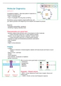
Translational medicine – Why does patient A respond to a
treatment and patient B not
- Fase 1: is the drug safe?
- Fase 2: Investigate if the drug does something
Sometimes a group of patients respond differently, and
you need to go back to the beginning of the research and repeat the process
Diagnosis
- Emergency presentation; symptoms
- Screening; Imaging, laboratory tests
Characteristics of a good test
- Sensitive; detect small amounts, even in the presence of other molecules
- -> Gives information about the % of false negative samples
- Specific; only the target molecule is detected = positive result
- -> Gives information about the % of false positive samples
- Potential for simple and standardized procedures (automation)
- High throughput
- Cheap
- Helps in clinical decisions
Imaging
- Location
- -> Based on interaction of electromagnetic radiation with body tissues and fluids or sound
waves
- Stage of tumor; (estimate)
- Growth
- Tumor dissection
- Plan treatment; (localization of radiation)
- Monitor recurrence
Why do some tumors grow faster or metastasize?
- Proliferation
- Angiogenesis
- Migration
- Apoptosis
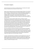
Example – Breast cancer
- HER2 = Human Epidermal Growth factor receptor (tissue and
cellular level)
- > HER2 = shorter survival, bad therapeutic response
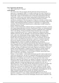
,FISH – Fluorescent In Situ Hybridization
- Presence of HER2
- Localize DNA sequences
- Control probes
- Monoclonal antibody; Herceptin (trastuzumab)
- -> Improved outcome of HER2 positive patients
Resistance to Herceptin treatment
- Unravel the mechanism; why do some women not respond to the treatment?
- Back to the bench
- Label antibodies to investigate problem
Intravital imaging
- Unravel why drugs fail; imaging of drug localization (incomplete tissue penetration by drug,
heterogeneous cell population, off-target)
- Hollow sphere / vesicle coupled to antibody
- -> Loaded with fluorescent dye
- -> Loaded with chemotherapeutic
- Fluorescent molecule coupled to antibody
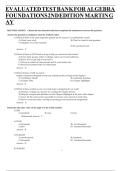
Studying drug delivery in vivo
- Animal models
- Functionalized molecules for
visualization
- Fluorophore couple to an antibody
- Vesicle coupled to an antibody
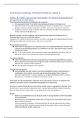
- In Vivo Imaging System – IVIS
- -> Tumor angiogenesis; visualize if
the drug inhibits angiogenesis
- Blue = nuclei
- Red = drug
- Green = Btk (brutons tyrosine kinase)
, Lecture 2
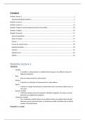
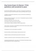
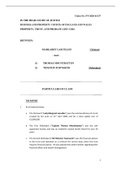
FRET – Fluorescent resonance energy
transfer
- Distance dependent energy transfer
- Donor – Acceptor
- Overlapping excitation/emission spectra
What is measured?
- Decrease of donor fluorescence
- Increase of acceptor fluorescence
2 molecules, containing
fluorescence. The donor
transfers energy to the
acceptor when they are in
close proximity.
Donor Acceptor
Emission = sending back light
in a different color - -> release
of energy in the form of light
The wavelength of the emission of the donor is the same as for the excitation of the acceptor.
- Can be used for studying protein – protein interactions
- Can be used for studying the structure of proteins (conformational changes) (drug
development)
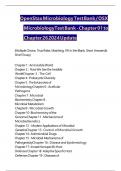
Chronic myeloid leukemia
- Study protein conformational changes with FRET
- Translocation of BCR to ABL = fusion protein
- Constitutively active tyrosine kinase - -> starts
phosphorylating, doesn’t stop
- -> Leads to phosphorylation to CrKL (good drug target,
for if you can prevent the phosphorylation)
- -> After the phosphorylation, the conformation changes
Phosphorylation - -> folding - -> energy transfer
- Drug target = BCR-ABL (tyrosine kinase)
- Novel drug = tyrosine kinase inhibitor - -> decreased fret
Lecture 1
Translational medicine – Why does patient A respond to a
treatment and patient B not
- Fase 1: is the drug safe?
- Fase 2: Investigate if the drug does something
Sometimes a group of patients respond differently, and
you need to go back to the beginning of the research and repeat the process
Diagnosis
- Emergency presentation; symptoms
- Screening; Imaging, laboratory tests
Characteristics of a good test
- Sensitive; detect small amounts, even in the presence of other molecules
- -> Gives information about the % of false negative samples
- Specific; only the target molecule is detected = positive result
- -> Gives information about the % of false positive samples
- Potential for simple and standardized procedures (automation)
- High throughput
- Cheap
- Helps in clinical decisions
Imaging
- Location
- -> Based on interaction of electromagnetic radiation with body tissues and fluids or sound
waves
- Stage of tumor; (estimate)
- Growth
- Tumor dissection
- Plan treatment; (localization of radiation)
- Monitor recurrence
Why do some tumors grow faster or metastasize?
- Proliferation
- Angiogenesis
- Migration
- Apoptosis
Example – Breast cancer
- HER2 = Human Epidermal Growth factor receptor (tissue and
cellular level)
- > HER2 = shorter survival, bad therapeutic response
,FISH – Fluorescent In Situ Hybridization
- Presence of HER2
- Localize DNA sequences
- Control probes
- Monoclonal antibody; Herceptin (trastuzumab)
- -> Improved outcome of HER2 positive patients
Resistance to Herceptin treatment
- Unravel the mechanism; why do some women not respond to the treatment?
- Back to the bench
- Label antibodies to investigate problem
Intravital imaging
- Unravel why drugs fail; imaging of drug localization (incomplete tissue penetration by drug,
heterogeneous cell population, off-target)
- Hollow sphere / vesicle coupled to antibody
- -> Loaded with fluorescent dye
- -> Loaded with chemotherapeutic
- Fluorescent molecule coupled to antibody
Studying drug delivery in vivo
- Animal models
- Functionalized molecules for
visualization
- Fluorophore couple to an antibody
- Vesicle coupled to an antibody
- In Vivo Imaging System – IVIS
- -> Tumor angiogenesis; visualize if
the drug inhibits angiogenesis
- Blue = nuclei
- Red = drug
- Green = Btk (brutons tyrosine kinase)
, Lecture 2
FRET – Fluorescent resonance energy
transfer
- Distance dependent energy transfer
- Donor – Acceptor
- Overlapping excitation/emission spectra
What is measured?
- Decrease of donor fluorescence
- Increase of acceptor fluorescence
2 molecules, containing
fluorescence. The donor
transfers energy to the
acceptor when they are in
close proximity.
Donor Acceptor
Emission = sending back light
in a different color - -> release
of energy in the form of light
The wavelength of the emission of the donor is the same as for the excitation of the acceptor.
- Can be used for studying protein – protein interactions
- Can be used for studying the structure of proteins (conformational changes) (drug
development)
Chronic myeloid leukemia
- Study protein conformational changes with FRET
- Translocation of BCR to ABL = fusion protein
- Constitutively active tyrosine kinase - -> starts
phosphorylating, doesn’t stop
- -> Leads to phosphorylation to CrKL (good drug target,
for if you can prevent the phosphorylation)
- -> After the phosphorylation, the conformation changes
Phosphorylation - -> folding - -> energy transfer
- Drug target = BCR-ABL (tyrosine kinase)
- Novel drug = tyrosine kinase inhibitor - -> decreased fret