Case 1 How is the brain organized? The start of new insights.
1. Macroscopic anatomy of the CNS, the brain and spinal cord:
Early development:
Two vesicles emerge from the prosencephalon: the telencephalon and the diencephalon. The mesencephalon
remains undivided throughout brain development, the rhombencephalon gives rise to the metencephalon and the
myelencephalon.
Central nervous system:
The central nervous system consists of the spinal cord and brain:
Spinal Cord:
- participates in the the transmission of both sensory information in the white matter axon tracts that ascend to
the brain and motor information in the descending tracts.
- Each spinal cord segment contains a pair of nerve roots called the dorsal and ventral roots.
- Dorsal roots contain sensory axons, which transmit sensory information into the spinal cord. Ventral roots
contain motor axons, which transmit motor commands to muscle and other body organs.
,Brainstem and cerebellum:
Consists of the medulla, the pons and the midbrain, it has 3 main functions:
- receiving sensory information from cranial structures and controlling the muscles of the head
- conducting information because ascending sensory and descending motor tracts travel through it.
- integrating diverse information from a variety of sources for arousal, behavioral responses to the environment,
and other higher brain functions.
- the principal functions of the cerebellum are to regulate eye and limb movements and to maintain posture and
balance.
Diencephalon:
Consists of the thalamus and hypothalamus:
Thalamus:
- transmits information to the cerebral hemispheres.
- is composed of several nuclei
Hypothalamus:
Controls endocrine hormone release from the pituitary gland and the overall functions of the autonomic nervous
system.
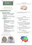
,Cerebral hemispheres:
Each brain hemisphere consists of: the cerebral cortex, hippocampal formation, amygdala, and basal ganglia.
The cerebral cortex is highly convoluted, the elevated convolutions are called gyri and are separated by grooves
called sulci or fissures (deep sulci). The cerebral hemispheres are separated from each other by the sagittal fissure.
Frontal lobe:
-serves diverse behavioral functions, from thoughts to action, cognition, and emotions.
- the precentral gyrus contains the primary motor cortex, which participates in controlling the mechanical actions
of movement. The premotor areas are adjacent to the primary motor cortex.
- the frontal lobe consists mainly of the association cortex.
Parietal lobe:
- is separated from the frontal lobe by the central sulcus.
- mediates perceptions of touch, pain, and limb position, carried out by the primary somatic sensory cortex
(located in the postcentral gyrus).
Occipital lobe:
-separated from the parietal lobe on the medial brain surface by the parietooccipital sulcus.
- the occipital lobe plays an important role in vision.
- the primary visual cortex is located in the walls of the calcarine fissure on the medial brain surface.
Temporal lobe:
- separated from the frontal and parietal lobes by the lateral sulcus (or Sylvian fissure)
- participates in memory and emotions
- consists of 3 parts: superior temporal gyrus, the middle temporal gyrus and inferior temporal gyrus.
, Insular lobe:
Deep in the lateral sulcus are portions of the frontal, parietal and temporal lobes called the insular cortex. The
insular cortex plays an important role in taste, internal body senses, pain and balance.
Corpus callosum:
The corpus callosum contains axons that interconnect the cortex of the two sides of the brain.
Cavities of CNS:
The ventricular system consists of cavities filled by cerebrospinal fluid located in the CNS. The cerebrospinal fluid is
a watery fluid secreted in the choroid plexus, it reduces physical shocks and plays and important role in chemical
communication.
1. Macroscopic anatomy of the CNS, the brain and spinal cord:
Early development:
Two vesicles emerge from the prosencephalon: the telencephalon and the diencephalon. The mesencephalon
remains undivided throughout brain development, the rhombencephalon gives rise to the metencephalon and the
myelencephalon.
Central nervous system:
The central nervous system consists of the spinal cord and brain:
Spinal Cord:
- participates in the the transmission of both sensory information in the white matter axon tracts that ascend to
the brain and motor information in the descending tracts.
- Each spinal cord segment contains a pair of nerve roots called the dorsal and ventral roots.
- Dorsal roots contain sensory axons, which transmit sensory information into the spinal cord. Ventral roots
contain motor axons, which transmit motor commands to muscle and other body organs.
,Brainstem and cerebellum:
Consists of the medulla, the pons and the midbrain, it has 3 main functions:
- receiving sensory information from cranial structures and controlling the muscles of the head
- conducting information because ascending sensory and descending motor tracts travel through it.
- integrating diverse information from a variety of sources for arousal, behavioral responses to the environment,
and other higher brain functions.
- the principal functions of the cerebellum are to regulate eye and limb movements and to maintain posture and
balance.
Diencephalon:
Consists of the thalamus and hypothalamus:
Thalamus:
- transmits information to the cerebral hemispheres.
- is composed of several nuclei
Hypothalamus:
Controls endocrine hormone release from the pituitary gland and the overall functions of the autonomic nervous
system.
,Cerebral hemispheres:
Each brain hemisphere consists of: the cerebral cortex, hippocampal formation, amygdala, and basal ganglia.
The cerebral cortex is highly convoluted, the elevated convolutions are called gyri and are separated by grooves
called sulci or fissures (deep sulci). The cerebral hemispheres are separated from each other by the sagittal fissure.
Frontal lobe:
-serves diverse behavioral functions, from thoughts to action, cognition, and emotions.
- the precentral gyrus contains the primary motor cortex, which participates in controlling the mechanical actions
of movement. The premotor areas are adjacent to the primary motor cortex.
- the frontal lobe consists mainly of the association cortex.
Parietal lobe:
- is separated from the frontal lobe by the central sulcus.
- mediates perceptions of touch, pain, and limb position, carried out by the primary somatic sensory cortex
(located in the postcentral gyrus).
Occipital lobe:
-separated from the parietal lobe on the medial brain surface by the parietooccipital sulcus.
- the occipital lobe plays an important role in vision.
- the primary visual cortex is located in the walls of the calcarine fissure on the medial brain surface.
Temporal lobe:
- separated from the frontal and parietal lobes by the lateral sulcus (or Sylvian fissure)
- participates in memory and emotions
- consists of 3 parts: superior temporal gyrus, the middle temporal gyrus and inferior temporal gyrus.
, Insular lobe:
Deep in the lateral sulcus are portions of the frontal, parietal and temporal lobes called the insular cortex. The
insular cortex plays an important role in taste, internal body senses, pain and balance.
Corpus callosum:
The corpus callosum contains axons that interconnect the cortex of the two sides of the brain.
Cavities of CNS:
The ventricular system consists of cavities filled by cerebrospinal fluid located in the CNS. The cerebrospinal fluid is
a watery fluid secreted in the choroid plexus, it reduces physical shocks and plays and important role in chemical
communication.