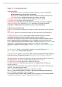
Chapter 19: The female genital system
Uterus has 3 layers:
- Endometrium; mucosa. Glandular structures and stroma. Tumors: endometrial
polyps (benign most) and endometrial carcinoma
- Myometrium; smooth muscle tissue. Tumors: leiomyomas (benign) and sarcomas
- serosa/peritoneum; mesothelial lining on the outside of the uterus
Leiomyoma: most common. Usually benign. No atypia, necrosis, or mitosis.
Leiomyosarcoma: see atypia, mitosis, and necrosis.
Endometrial polyp: risk factors by obesity and use of tamoxifen. Can become malignant.
Atypical hyperplasia (EIN): long term oestrogen stimulation. Increased ratio glands.
Inactivation of PTEN. High risk of endometrial cancer. In the endometrium.
Endometriosis and adenomyosis:
Endometriosis: presence of endometrium outside the uterus. Also called ovarian chocolate
cyst.
Adenomyosis: presence of endometrium inside the uterus but then in the myometrium.
Endometrioid adenocarcinoma: most common (80%) malignant tumor in the uterus.
Serous adenocarcinoma and Clear cell adenocarcinoma are less common.
Risk factors are oestrogen exposure, less common is germline mutation. Clinical course by
post menopause bleeding. Mostly diagnosed with low stage. Survival state depends on
grade. Endometrioid adenocarcinoma has 3 grades, depending on their differentiation. Most
times grade 1. When grading increases, irregularity increases with severe atypia.
Hyperplasia background. Staining pattern is PAX8+, P53 wildtype, loss of PTEN, P16 patchy,
ER+.
Serous carcinoma: Grade 3. Loss of polarity. Happens in atrophic background. P53++, P16+,
WT1+ = ovarian origin. Most common in ovaries.
Clear cell carcinoma: grade 3. Less common than serous. Also papillary formation. Nucleus is
projected in lumen = hobnailing. Staining by: P16 patchy, ER & PR negative, Napasine A
HPV related cancer: cervix but also other types like anus, vulva, penis. High risk on cancer in
the types 16, 18, 31, 33 and 45. Cervical cancer most times P16+ in HPV.
Productive infection: active replication of DNA of the HPV virus
Transforming infection: sometimes some genes are not regulated well, and those can
become cancerous.
Happens often in transformation zone. Squamous epithelium on outside, and inside uterus
columnar. Here often precursor lesion.
CIN 1 = cervical intraepithelial neoplasia. Low grade. P16- or patchy
CIN 2 & 3 = high grade neoplasia. The extent of the disturbed epithelium determine the
grade. P16 higher staining depending on the grade.
The screening goes via the HPV. When positive, further screening. You want glandular cells
and squamous cells, then it is a good scrape of transformation zone.