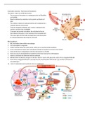
Haemolytic anaemias - Red blood cell breakdown
The spleen: major role in RBC destruction
- The structure of the spleen is a testing ground of cell flexibility
and viability
- Blood is delivered by arterioles to the splenic red blood cell
pulp
- The volume of plasma is reduced and the cell is subjected to a
relatively hypoxic environment
- Tests the metabolic pathways and, in older or diseased cells,
results in a further loss of pliability
- To escape and re-enter circulation, the red blood cell must
then squeeze through a 2-5 μm opening in the sinusoidal wall
- This traps rigid cells and leads to phagocytosis and destruction
by reticuloendothelial cells lining the sinusoids
RBC breakdown:
1. RBC are broken down within macrophages
2. The haemoglobin is degraded
3. Globin is broken down into amino acids, which are re-used for protein synthesis
4. Iron is removed from haem and in the plasma it binds to transferrin in its ferric form (Fe3+).
5. Some of this iron is transported to the liver and stored in as Ferritin
6. Some iron may be transported by transferrin to the bone marrow for erythropoiesis
7. Haem is converted into biliverdin and then bilirubin.
8. Bilirubin binds to albumin and goes to the liver where it reacts with glucuronic acid to form conjugated bilirubin
9. Most of the conjugated bilirubin is secreted into the small intestine with the bile (12) and then converted to
stercobilinogen.
10. Some is reabsorbed and excreted in urine as urobilinogen
, RBC breakdown: intravascular:
- RBCs are destroyed within the vascular compartment
- Plays only a small part of normal red cell destruction
- Haemoglobin (Hb) is released and is rapidly bound by the serum protein haptoglobin
- The haptoglobin-Hb complex is recognised by monocytes/macrophages and degraded
- Formation of large amounts of haptoglobin-Hb complexes leads to rapid haptoglobin depletion
- Haem in Fe3+ state binds to albumin to become methemalbumin
- Excess free Hb goes to kidney and excreted in urine (haemoglobinuria) – hemosiderinuria (brown
urine) also occurs due to iron accumulation in renal tubules
Introduction to haemolytic anaemia:
- Results from an increase in the rate of RBC destruction
- Due to increased RBC destruction, the bone marrow presents hyperplasia, this means that it will produce RBCs at
6-8 times the normal rate
- Therefore may not see haemolytic anaemia until lifespan of red cells <30 days
- The reticulocytes count is normally increased because the increased production of RBCs
Classification of haemolytic anaemias:
Symptoms and presentation:
1. Typical symptoms of anaemia:
a. Fatigue