1
Ophthalmology
History and examination......................................................................................................................... 2
Anatomy ................................................................................................................................................ 4
Presentations ......................................................................................................................................... 8
Vision loss ........................................................................................................................................................ 8
Abnormal pupil ................................................................................................................................................ 8
Colour Blindness ............................................................................................................................................ 10
Red Eye .......................................................................................................................................................... 10
Diplopia ......................................................................................................................................................... 12
Emergencies ................................................................................................................................................... 14
Chemical injuries ...................................................................................................................................................................... 15
Deterrant spray injuries ........................................................................................................................................................... 15
Penetrating injuries .................................................................................................................................................................. 15
Orbital blowout fracture .......................................................................................................................................................... 16
Cavernous sinus thrombosis .................................................................................................................................................... 16
Corneal abrasion ...................................................................................................................................................................... 17
Corneal ulcer (infective keratitis) ............................................................................................................................................. 17
Optic neuritis ............................................................................................................................................................................ 17
Cataracts .............................................................................................................................................. 18
Ectopia Lentis ....................................................................................................................................... 19
Hypertensive Retinopathy..................................................................................................................... 19
Diabetic Retinopathy ............................................................................................................................ 19
Conjunctivitis (Pink Eye) ........................................................................................................................ 21
Subconjunctival haemorrhage .............................................................................................................. 22
Iritis (Anterior Uveitis or Iridocyclitis) .................................................................................................... 22
Uveitis (Posterior Uveitis or Choroiditis) ................................................................................................ 23
Corneal Problems ................................................................................................................................. 24
Scleritis................................................................................................................................................. 26
Episcleritis ............................................................................................................................................ 27
Glaucoma ............................................................................................................................................. 28
Primary Open-angle Glaucoma ....................................................................................................................... 29
Closed-angle/Acute Angle-Closure Glaucoma (EMERGENCY) ........................................................................... 30
Periorbital/Pre-Septal Cellulitis ............................................................................................................. 31
Orbital/Post-Septal Cellulitis ................................................................................................................. 31
Eyelid Problems .................................................................................................................................... 32
Stye ............................................................................................................................................................... 32
Chalazion/Meibomian Cyst ............................................................................................................................. 33
, 2
Blepharitis ..................................................................................................................................................... 33
Abnormal Eyelid Position................................................................................................................................ 33
Retinal Vascular Occlusions .................................................................................................................. 35
Anterior ischaemic optic neuropathy .............................................................................................................. 35
Central Retinal Vein Occlusion ........................................................................................................................ 36
Central Retinal Artery Occlusion ..................................................................................................................... 36
Retinal Detachment .............................................................................................................................. 37
Cranial Nerve Palsies and the Eye ......................................................................................................... 40
Refraction and Refractive Errors ........................................................................................................... 41
Squint (Strabismus)............................................................................................................................... 43
Thyroid Eye Disease (Grave’s Orbitopathy) ............................................................................................ 45
History and examination
History:
• Onset
• Trauma?
• Location – unilateral, bilateral, sectoral
• Pain/discomfort (gritty, FB sensation, itch, deep ache)
o FB sensation – FB, conjunctivitis, blepharitis, abrasion or ulcer
o Gritty – corneal ulcer, corneal abrasion, FB
o Lid itching – blepharitis, stye, chalazion, eczema
o Aching in eyeball – scleritis or uveitis
o Pressure sensation with N+V – acute angle closure glaucoma
o Headache – migraine, meningitis, encephalitis, cluster headache, ICP, GCA
o Eye pain – inside the eye or outside?
• Photosensitivity/photophobia
• Redness (one/both eyes, diffuse, perilimbal, sectoral, colour – bright or dark red, vessel dilation or
haemorrhage)
• Watering +/- discharge Photopsia –
• Change in vision (blurring, halos around lights, diplopia, photopsia) flashing lights
o Blurring – generalised or central?
▪ Generalised blurring – reduction in clarity of entire field of vision. Caused by refractive error,
cataracts (diffuse, painless loss of vision), medication SEs e.g. steroids
▪ Central blurring – pathology more likely to be in the macula or optic nerve. Caused by age
related macula degeneration, diabetic maculopathy, optic disc swelling
o Floaters (blob moves about), it’s in the vitreous humour e.g. posterior vitreous detachment, vitritis,
blood
o Scotoma (blob stays in one place) - it could mean retina detachment or vascular occlusion
o Photopsia – usually due to migraine (Sx go after aura ends) or pathology in vitreous or retina
(vitreous detachment, retinal tear or detachment)
o Diplopia – clarify what they mean by double vision. Ask when they cover one eye, if one image
disappears then they have monocular diplopia
▪ Monocular – double vision is present even if one eye is closed if you cover up the normal
eye. If you cover up the eye with double vision, only one image will show and double vision
will no longer be present. This is due to cataracts, corneal abrasion, eyelid cyst
, 3
▪ Binocular – double vision is only present when both eyes are open. This is due to pathology
In the nerves, NMJ or muscles
• Contact lens wear/ glasses (ask if they have glasses for reading/driving/TV etc)
• Previous ocular history
• PMH
Examination:
• Inspect face, eyelids and area around the eyes
o Inspect periocular region for abnormalities
o Ocular adnexa – area around the eye
• Conjunctiva – check for:
o Congestion – redness (diffuse)
o Subconjunctival haemorrhage
o Discolouration – yellowing (jaundice)
• Cornea – check for:
o Normal transparency
o Haze
o Oedema
o Cloudiness
• Anterior chamber – check for:
o Hyphema – blood in anterior chamber. Causes include clotting problems and trauma
• Iris – check against the other eye:
o Haemorrhage
• Pupil – check against the other eye:
o Size
o Shape
o Reaction – light reaction and accommodation reflex
Posterior segment requires gadgets to test for abnormalities.
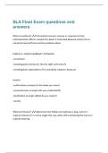
Visual Acuity Tests – Snellen chart (uses score of 6/6), LogMAR chart (uses score of 0.1 per line with each letter
having value of 0.02, goes down from 1.0 [6/60] to 0.0 [6/6] with better visual acuity).
• 6/12 or 0.3 is minimum driving standard by DVLA
• Children:
o Keeler Preferential Looking Cards – 8wks-12mths, uses square-wave grating of different spatial
frequencies with examiner objectively observing patient’s eyes to see if look toward grating
o Cardiff Acuity Cards – 3-18mths, simple picture at top of bottom, check if eyes looking towards top
or bottom of card
o Kay Pictures – 2-4yrs, picture-based test +/- matching cards, recorded as LogMAR, 4 pics each line at
3m
o LogMAR Keeler Book – 4yrs+, letter-based +/- matching cards, test at 3m
o Testing Low Vision – counting fingers, hand movements, perception of light, no perception of light
(if cannot perceive light)
, 4
Other Visual Tests – Ishihara colour vision test
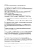
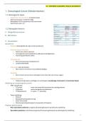
Supraorbital nerve,
artery and vein
Anatomy
Orbital cavity – made of 4 bony walls. Cavity is a pyramidal
shape with the apex facing anteriorly.
Blood vessels and nerves of orbit – main arterial supply is via
CNIII, CNIV, CNVI ophthalmic artery and its branches. Ophthalmic veins
(superior and inferior) drain venous blood into cavernous
Maxillary
nerve,
infra-
orbital
nerve/art
ery/vein,
zygomatic
nerve,
ganglionic
branches
from Infraorbital branch of
pterygop maxillary division - CNV
alatine
ganglion
sinus, pterygoid plexus and facial vein. General sensory
innervation from the eye is via ophthalmic division of
trigeminal (Va). Special sensory vision from retina is
transmitted via the optic nerve. Motor innervation to
muscles is via CN III, IV, VI.
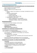
Eyelid – consists of skin, subcutaneous tissue, tarsal plate and muscles (palpebral part of orbicularis oculi and levator
palpebrae superioris). Also consist of glands (Meibomian and sebaceous glands that are associated with lash follicle).
Meibomian glands secrete a lipid-rich substance onto edges of lid to prevent evaporation of tear film and tear
spillage.
Ophthalmology
History and examination......................................................................................................................... 2
Anatomy ................................................................................................................................................ 4
Presentations ......................................................................................................................................... 8
Vision loss ........................................................................................................................................................ 8
Abnormal pupil ................................................................................................................................................ 8
Colour Blindness ............................................................................................................................................ 10
Red Eye .......................................................................................................................................................... 10
Diplopia ......................................................................................................................................................... 12
Emergencies ................................................................................................................................................... 14
Chemical injuries ...................................................................................................................................................................... 15
Deterrant spray injuries ........................................................................................................................................................... 15
Penetrating injuries .................................................................................................................................................................. 15
Orbital blowout fracture .......................................................................................................................................................... 16
Cavernous sinus thrombosis .................................................................................................................................................... 16
Corneal abrasion ...................................................................................................................................................................... 17
Corneal ulcer (infective keratitis) ............................................................................................................................................. 17
Optic neuritis ............................................................................................................................................................................ 17
Cataracts .............................................................................................................................................. 18
Ectopia Lentis ....................................................................................................................................... 19
Hypertensive Retinopathy..................................................................................................................... 19
Diabetic Retinopathy ............................................................................................................................ 19
Conjunctivitis (Pink Eye) ........................................................................................................................ 21
Subconjunctival haemorrhage .............................................................................................................. 22
Iritis (Anterior Uveitis or Iridocyclitis) .................................................................................................... 22
Uveitis (Posterior Uveitis or Choroiditis) ................................................................................................ 23
Corneal Problems ................................................................................................................................. 24
Scleritis................................................................................................................................................. 26
Episcleritis ............................................................................................................................................ 27
Glaucoma ............................................................................................................................................. 28
Primary Open-angle Glaucoma ....................................................................................................................... 29
Closed-angle/Acute Angle-Closure Glaucoma (EMERGENCY) ........................................................................... 30
Periorbital/Pre-Septal Cellulitis ............................................................................................................. 31
Orbital/Post-Septal Cellulitis ................................................................................................................. 31
Eyelid Problems .................................................................................................................................... 32
Stye ............................................................................................................................................................... 32
Chalazion/Meibomian Cyst ............................................................................................................................. 33
, 2
Blepharitis ..................................................................................................................................................... 33
Abnormal Eyelid Position................................................................................................................................ 33
Retinal Vascular Occlusions .................................................................................................................. 35
Anterior ischaemic optic neuropathy .............................................................................................................. 35
Central Retinal Vein Occlusion ........................................................................................................................ 36
Central Retinal Artery Occlusion ..................................................................................................................... 36
Retinal Detachment .............................................................................................................................. 37
Cranial Nerve Palsies and the Eye ......................................................................................................... 40
Refraction and Refractive Errors ........................................................................................................... 41
Squint (Strabismus)............................................................................................................................... 43
Thyroid Eye Disease (Grave’s Orbitopathy) ............................................................................................ 45
History and examination
History:
• Onset
• Trauma?
• Location – unilateral, bilateral, sectoral
• Pain/discomfort (gritty, FB sensation, itch, deep ache)
o FB sensation – FB, conjunctivitis, blepharitis, abrasion or ulcer
o Gritty – corneal ulcer, corneal abrasion, FB
o Lid itching – blepharitis, stye, chalazion, eczema
o Aching in eyeball – scleritis or uveitis
o Pressure sensation with N+V – acute angle closure glaucoma
o Headache – migraine, meningitis, encephalitis, cluster headache, ICP, GCA
o Eye pain – inside the eye or outside?
• Photosensitivity/photophobia
• Redness (one/both eyes, diffuse, perilimbal, sectoral, colour – bright or dark red, vessel dilation or
haemorrhage)
• Watering +/- discharge Photopsia –
• Change in vision (blurring, halos around lights, diplopia, photopsia) flashing lights
o Blurring – generalised or central?
▪ Generalised blurring – reduction in clarity of entire field of vision. Caused by refractive error,
cataracts (diffuse, painless loss of vision), medication SEs e.g. steroids
▪ Central blurring – pathology more likely to be in the macula or optic nerve. Caused by age
related macula degeneration, diabetic maculopathy, optic disc swelling
o Floaters (blob moves about), it’s in the vitreous humour e.g. posterior vitreous detachment, vitritis,
blood
o Scotoma (blob stays in one place) - it could mean retina detachment or vascular occlusion
o Photopsia – usually due to migraine (Sx go after aura ends) or pathology in vitreous or retina
(vitreous detachment, retinal tear or detachment)
o Diplopia – clarify what they mean by double vision. Ask when they cover one eye, if one image
disappears then they have monocular diplopia
▪ Monocular – double vision is present even if one eye is closed if you cover up the normal
eye. If you cover up the eye with double vision, only one image will show and double vision
will no longer be present. This is due to cataracts, corneal abrasion, eyelid cyst
, 3
▪ Binocular – double vision is only present when both eyes are open. This is due to pathology
In the nerves, NMJ or muscles
• Contact lens wear/ glasses (ask if they have glasses for reading/driving/TV etc)
• Previous ocular history
• PMH
Examination:
• Inspect face, eyelids and area around the eyes
o Inspect periocular region for abnormalities
o Ocular adnexa – area around the eye
• Conjunctiva – check for:
o Congestion – redness (diffuse)
o Subconjunctival haemorrhage
o Discolouration – yellowing (jaundice)
• Cornea – check for:
o Normal transparency
o Haze
o Oedema
o Cloudiness
• Anterior chamber – check for:
o Hyphema – blood in anterior chamber. Causes include clotting problems and trauma
• Iris – check against the other eye:
o Haemorrhage
• Pupil – check against the other eye:
o Size
o Shape
o Reaction – light reaction and accommodation reflex
Posterior segment requires gadgets to test for abnormalities.
Visual Acuity Tests – Snellen chart (uses score of 6/6), LogMAR chart (uses score of 0.1 per line with each letter
having value of 0.02, goes down from 1.0 [6/60] to 0.0 [6/6] with better visual acuity).
• 6/12 or 0.3 is minimum driving standard by DVLA
• Children:
o Keeler Preferential Looking Cards – 8wks-12mths, uses square-wave grating of different spatial
frequencies with examiner objectively observing patient’s eyes to see if look toward grating
o Cardiff Acuity Cards – 3-18mths, simple picture at top of bottom, check if eyes looking towards top
or bottom of card
o Kay Pictures – 2-4yrs, picture-based test +/- matching cards, recorded as LogMAR, 4 pics each line at
3m
o LogMAR Keeler Book – 4yrs+, letter-based +/- matching cards, test at 3m
o Testing Low Vision – counting fingers, hand movements, perception of light, no perception of light
(if cannot perceive light)
, 4
Other Visual Tests – Ishihara colour vision test
Supraorbital nerve,
artery and vein
Anatomy
Orbital cavity – made of 4 bony walls. Cavity is a pyramidal
shape with the apex facing anteriorly.
Blood vessels and nerves of orbit – main arterial supply is via
CNIII, CNIV, CNVI ophthalmic artery and its branches. Ophthalmic veins
(superior and inferior) drain venous blood into cavernous
Maxillary
nerve,
infra-
orbital
nerve/art
ery/vein,
zygomatic
nerve,
ganglionic
branches
from Infraorbital branch of
pterygop maxillary division - CNV
alatine
ganglion
sinus, pterygoid plexus and facial vein. General sensory
innervation from the eye is via ophthalmic division of
trigeminal (Va). Special sensory vision from retina is
transmitted via the optic nerve. Motor innervation to
muscles is via CN III, IV, VI.
Eyelid – consists of skin, subcutaneous tissue, tarsal plate and muscles (palpebral part of orbicularis oculi and levator
palpebrae superioris). Also consist of glands (Meibomian and sebaceous glands that are associated with lash follicle).
Meibomian glands secrete a lipid-rich substance onto edges of lid to prevent evaporation of tear film and tear
spillage.