Human anatomy and physiology
Lecture 1 – Anatomy of the heart
- What is the anatomical position of the heart in the thorax? sternal angle, 5th intercostal
space: parasternal on the right, midclavicular on the left
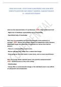
- What is the mediastinum? space within the rib cage between the lung cavities
- What is the structure of the heart?
Sternal angle: base of heart
Intercostal: between ribs
Parasternal: next to the sternum
Midclavicular: mid of “sleutelbeen”
Mediastinum:
- Superior mediastinum above the heart
1. Thymus lymphoid tissue, regression
2. Nerves
Vagal nerves: regulation of internal organ functions
Left recurrent laryngeal nerve: control intrinsic muscles of the larynx (except cricothyroid
muscle)
Phrenic nerves: causes hiccups, enters the diaphragm (major muscle of respiration,
located below the lungs)
Sympathetic trunk: component of the autonomic nervous system
3. Thoracic duct lymphatic drainage
4. Vessels aa. and vv. thoracicae internae (internal thoracic arteries)
- Inferior mediastinum contains the heart
1. Anterior space between the sternum and the heart
2. Middle the heart
3. Posterior descending aorta, esophagus, vessels, thoracic duct, nerves (vagal nerves,
sympathetic trunk)
Pericardial cavity: balloon in which everything moves smoothly, fluid causes cardiac tamponade
- Visceral pleura membrane that covers each lung
- Parietal membrane that is attached to the thoracic (or other body) cavity
Transverse sinus(/cavity): transition from visceral to parietal
Oblique sinus:
Coronary sinus: collects blood from the heart (independent circulation)
Interventricular sulcus: 2 grooves (anterior and posterior) that separates the
ventricles of the heart
When parietal cavity is punctured fills with blood; heart cannot expand
Trunk + anatomical context it will split at some point
E.g., pulmonary trunk: 2 pulmonary arteries
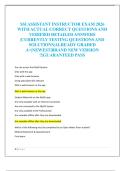
Right atrium (posterior):
- Right auricula (pectinate muscles)
- Vena cava (inferior + superior)
- Coronary sinus
, - Interatrial septum (oval fossa)
- Crista terminalis ridge between the smooth and rough wall
Right ventricle (anterior):
- Tricuspid valve septal, anterior, posterior cusps; chordae tendinae, papillary muscles
- Pulmonary valve (semilunar)
- Pulmonary trunk
Left atrium:
- Left auricle
- Pulmonary veins
- Interatrial septum
Left ventricle:
- Thicker wall greater circulation to entire body
- Ascending aorta
- Aortic valve (semilunar)
- Left atrioventricular valve (mitral, bicuspid)
What do valves need to function? firm connective tissue around the valves, arteries
Diastole relaxation of the heart after contraction
Systole contraction of the ventricles to pump blood
Heart receives blood during relaxation (diastole)
Conduction system:
- SA node gives off rhythm without brain interference (own rhythm)
- AV node stops the signal and gives off signal after x milliseconds (own rhythm)
- Bundle of His bundle branches
- Purkinje fibers
4 heart valves:
- Tricuspid valve between the right atrium and the right ventricle
- Pulmonary valve between the right ventricle and the pulmonary artery
- Mitral valve between the left atrium and the left ventricle
- Aortic valve between the left ventricle and the aorta
, Lecture 2 – Embryology of the heart
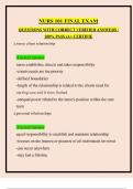
Heart formation:
1. Forming straight heart tube lateral heart tubes form together to form one tube
- Venous pole blood comes in, up)
- Arterial pole blood comes out, down (dorsal aorta)
2. Forming primitive heart
- Looping atrium migrates to cranial (up)
- Rotation right atrium and ventricle rotate to ventral
Pericardium development:
1. Primordial heart tube in pericardial sac
2. Primordial transverse pericardial sinus
3. Heart loops ventrally
4. Primordial arterial and venous poles form transverse pericardial sinus
5. Veins expansion + pericardial reflection form oblique pericardial sinus
3. Forming fetal heart
1. Septation atria septum/foramen 1primum and secundum foramen ovale
2. Septation ventricles
4. Post-natal changes
Situs inversus malformation in which the apex is on the right (dextrocardia)
Ventricular septum defect last step; therefore the most common defect
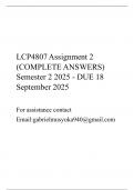
Tetralogy of Fallot (4 things happening):
1. Pulmonary stenosis (narrowing)
2. VSD (ventricular septum defect) right ventricle has to work harder to pump blood into
narrow pulmonary artery
3. Blood gets pumped into the aorta overriding aorta
4. Enlarged right ventricle
Lecture 1 – Anatomy of the heart
- What is the anatomical position of the heart in the thorax? sternal angle, 5th intercostal
space: parasternal on the right, midclavicular on the left
- What is the mediastinum? space within the rib cage between the lung cavities
- What is the structure of the heart?
Sternal angle: base of heart
Intercostal: between ribs
Parasternal: next to the sternum
Midclavicular: mid of “sleutelbeen”
Mediastinum:
- Superior mediastinum above the heart
1. Thymus lymphoid tissue, regression
2. Nerves
Vagal nerves: regulation of internal organ functions
Left recurrent laryngeal nerve: control intrinsic muscles of the larynx (except cricothyroid
muscle)
Phrenic nerves: causes hiccups, enters the diaphragm (major muscle of respiration,
located below the lungs)
Sympathetic trunk: component of the autonomic nervous system
3. Thoracic duct lymphatic drainage
4. Vessels aa. and vv. thoracicae internae (internal thoracic arteries)
- Inferior mediastinum contains the heart
1. Anterior space between the sternum and the heart
2. Middle the heart
3. Posterior descending aorta, esophagus, vessels, thoracic duct, nerves (vagal nerves,
sympathetic trunk)
Pericardial cavity: balloon in which everything moves smoothly, fluid causes cardiac tamponade
- Visceral pleura membrane that covers each lung
- Parietal membrane that is attached to the thoracic (or other body) cavity
Transverse sinus(/cavity): transition from visceral to parietal
Oblique sinus:
Coronary sinus: collects blood from the heart (independent circulation)
Interventricular sulcus: 2 grooves (anterior and posterior) that separates the
ventricles of the heart
When parietal cavity is punctured fills with blood; heart cannot expand
Trunk + anatomical context it will split at some point
E.g., pulmonary trunk: 2 pulmonary arteries
Right atrium (posterior):
- Right auricula (pectinate muscles)
- Vena cava (inferior + superior)
- Coronary sinus
, - Interatrial septum (oval fossa)
- Crista terminalis ridge between the smooth and rough wall
Right ventricle (anterior):
- Tricuspid valve septal, anterior, posterior cusps; chordae tendinae, papillary muscles
- Pulmonary valve (semilunar)
- Pulmonary trunk
Left atrium:
- Left auricle
- Pulmonary veins
- Interatrial septum
Left ventricle:
- Thicker wall greater circulation to entire body
- Ascending aorta
- Aortic valve (semilunar)
- Left atrioventricular valve (mitral, bicuspid)
What do valves need to function? firm connective tissue around the valves, arteries
Diastole relaxation of the heart after contraction
Systole contraction of the ventricles to pump blood
Heart receives blood during relaxation (diastole)
Conduction system:
- SA node gives off rhythm without brain interference (own rhythm)
- AV node stops the signal and gives off signal after x milliseconds (own rhythm)
- Bundle of His bundle branches
- Purkinje fibers
4 heart valves:
- Tricuspid valve between the right atrium and the right ventricle
- Pulmonary valve between the right ventricle and the pulmonary artery
- Mitral valve between the left atrium and the left ventricle
- Aortic valve between the left ventricle and the aorta
, Lecture 2 – Embryology of the heart
Heart formation:
1. Forming straight heart tube lateral heart tubes form together to form one tube
- Venous pole blood comes in, up)
- Arterial pole blood comes out, down (dorsal aorta)
2. Forming primitive heart
- Looping atrium migrates to cranial (up)
- Rotation right atrium and ventricle rotate to ventral
Pericardium development:
1. Primordial heart tube in pericardial sac
2. Primordial transverse pericardial sinus
3. Heart loops ventrally
4. Primordial arterial and venous poles form transverse pericardial sinus
5. Veins expansion + pericardial reflection form oblique pericardial sinus
3. Forming fetal heart
1. Septation atria septum/foramen 1primum and secundum foramen ovale
2. Septation ventricles
4. Post-natal changes
Situs inversus malformation in which the apex is on the right (dextrocardia)
Ventricular septum defect last step; therefore the most common defect
Tetralogy of Fallot (4 things happening):
1. Pulmonary stenosis (narrowing)
2. VSD (ventricular septum defect) right ventricle has to work harder to pump blood into
narrow pulmonary artery
3. Blood gets pumped into the aorta overriding aorta
4. Enlarged right ventricle