Histology: Contractile Cells
Enable movement of the body and movement within the body by contraction
Most completed by the protein’s actin and myosin
Single cell contraction units
1) myofibroblasts – contract and secrete collagen. Present in the process of
healing and repair, leading to the formation of a scar
2) Pericytes – surround blood vessels
3) Myoepithelial – found in secretory glands
Multicellular contraction units (muscle)
1) Skeletal muscle (voluntary)
2) Cardiac muscle (involuntary)
3) Smooth muscle (involuntary)
Skeletal muscle
Responsible for the movement of the skeleton, the orbit of the eye and the tongue
Striated appearance actin and myosin are arranged in a striated way
Cytoplasmic organelles have highly developed functions so are given different names
Plasma/cell membrane = sarcolemma
Cytoplasm = sarcoplasm
Endoplasmic reticulum = sarcoplasmic reticulum
Skeletal muscle is formed during embryogenesis with precursor cells fusing myoblasts
Makes skeletal muscle multinucleate (each skeletal muscle could have several nuclei
due to the fusing of the precursor cells)
Nuclei appear just below the sarcolemma
Surrounded by an external lamina
In addition to actin and myosin
Mitochondria and glycogen produce energy
Each cell is surrounded by and external lamina
In adults the precursor cells are present (satellite cells)
Satellite cells take over repairing muscle when it is damaged to that the muscle
can continue to function at an efficient level
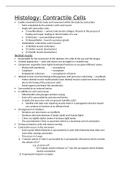
Arrangement of striations
Striations are also known as myofibrils
Striations alternate between A bands (dark) and I bands (light)
Z lines are slightly darker banks in between light bands
The area between Z lines is sarcomere which is a functional unit of contraction
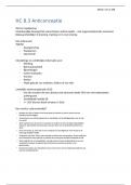
During contraction actin and myosin overlap
Contraction occurs in the sarcomere
Each myosin (thick) filament is surrounded by 6 actin (thin) filaments that slide over
each other causing contraction
This occurs in 2 ways
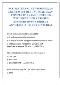
1) T tubular system (T tube is surrounded by 2 sarcoplasmic reticulums) which controls
the release of Ca2+
e.g. a) nerve cell
b) T tubular system releases Ca2+ into the sarcoplasm which initiates
muscle contraction
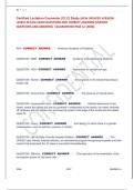
2) Tropomyosin/troponin complex
, Tropomyosin winds around actin to stabilise and to stiffen it
The troponin complex, which regulates the binding of actin to myosin, is attached
to tropomyosin and masks actin binding sites
Ca2+ released by the T tubular system is then bound to the troponin causing a
configurational change in troponin
Unmasks the binding site on actin
Myosin can then bind to the actin filament
Actin and myosin bind and slide over one another = a sliding filament mechanism
resulting in contraction
Abnormalities in skeletal muscle
Hypertrophy: an increase in muscle size not in muscle cell number
Atrophy: a decrease in muscle size not in muscle cell number
Often found in wheelchair/bed bound patients
Myasthenia Gravis: when nerve impulses are not transmitted effectively to the muscle;
the patient experiences weakness and rapid muscle fatigue upon voluntary movement
e.g. walking running
Muscular dystrophy: weakness and wasting of muscles due to a defect in one of the
proteins involved in muscle function
Fibres undergo progressive damage with repeated contraction, ultimately leading to
death of the muscle cells
Enable movement of the body and movement within the body by contraction
Most completed by the protein’s actin and myosin
Single cell contraction units
1) myofibroblasts – contract and secrete collagen. Present in the process of
healing and repair, leading to the formation of a scar
2) Pericytes – surround blood vessels
3) Myoepithelial – found in secretory glands
Multicellular contraction units (muscle)
1) Skeletal muscle (voluntary)
2) Cardiac muscle (involuntary)
3) Smooth muscle (involuntary)
Skeletal muscle
Responsible for the movement of the skeleton, the orbit of the eye and the tongue
Striated appearance actin and myosin are arranged in a striated way
Cytoplasmic organelles have highly developed functions so are given different names
Plasma/cell membrane = sarcolemma
Cytoplasm = sarcoplasm
Endoplasmic reticulum = sarcoplasmic reticulum
Skeletal muscle is formed during embryogenesis with precursor cells fusing myoblasts
Makes skeletal muscle multinucleate (each skeletal muscle could have several nuclei
due to the fusing of the precursor cells)
Nuclei appear just below the sarcolemma
Surrounded by an external lamina
In addition to actin and myosin
Mitochondria and glycogen produce energy
Each cell is surrounded by and external lamina
In adults the precursor cells are present (satellite cells)
Satellite cells take over repairing muscle when it is damaged to that the muscle
can continue to function at an efficient level
Arrangement of striations
Striations are also known as myofibrils
Striations alternate between A bands (dark) and I bands (light)
Z lines are slightly darker banks in between light bands
The area between Z lines is sarcomere which is a functional unit of contraction
During contraction actin and myosin overlap
Contraction occurs in the sarcomere
Each myosin (thick) filament is surrounded by 6 actin (thin) filaments that slide over
each other causing contraction
This occurs in 2 ways
1) T tubular system (T tube is surrounded by 2 sarcoplasmic reticulums) which controls
the release of Ca2+
e.g. a) nerve cell
b) T tubular system releases Ca2+ into the sarcoplasm which initiates
muscle contraction
2) Tropomyosin/troponin complex
, Tropomyosin winds around actin to stabilise and to stiffen it
The troponin complex, which regulates the binding of actin to myosin, is attached
to tropomyosin and masks actin binding sites
Ca2+ released by the T tubular system is then bound to the troponin causing a
configurational change in troponin
Unmasks the binding site on actin
Myosin can then bind to the actin filament
Actin and myosin bind and slide over one another = a sliding filament mechanism
resulting in contraction
Abnormalities in skeletal muscle
Hypertrophy: an increase in muscle size not in muscle cell number
Atrophy: a decrease in muscle size not in muscle cell number
Often found in wheelchair/bed bound patients
Myasthenia Gravis: when nerve impulses are not transmitted effectively to the muscle;
the patient experiences weakness and rapid muscle fatigue upon voluntary movement
e.g. walking running
Muscular dystrophy: weakness and wasting of muscles due to a defect in one of the
proteins involved in muscle function
Fibres undergo progressive damage with repeated contraction, ultimately leading to
death of the muscle cells