Transport in Mammals
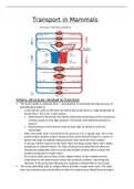
Artery structure related to function
The tunica media is relatively thick well adapted to withstand the high pressure of
blood flowing within it
In the arteries close to the heart (as well as the aorta) there is a high proportion of
elastic fibres. This is for 2 main reason:
When blood is forced into the arteries following contractions of the ventricles it
created a pulse of very high pressure. The elastic walls allow the arteries to
expand
Blood pressure in the arteries must be kept high for blood to reach the
extremities
When the elastic wall is stretched by the pressure in it, it springs back. This recoil
action creates another surge of pressure that carries blood forward in a series of
pulses and helps to maintain blood pressure even when the heart relaxes
In arteries further away from the heart there are fewer elastic fibres and a higher
proportion of smooth muscle. The flow of blood is less pulse-like but does not
smooth out completely until it reaches the smallest arteries which contain the
smallest proportion of elastic fibres
The tunica media also contains some collagen fibres. In the smallest arteries
contraction of the small muscle causes the vessels to constrict, narrowing the
diameter of the lumen and allowing the regulation of blood flow to the tissues
The tunica adventitia with its collagen fibres provides a tough outer layer. This outer
layer also contains some elastic fibres to allow for stretching as blood flows through it
, The overall thickness of the wall is large. This helps prevent arteries bursting under
pressure. Arteries have a relatively small lumen in proportion to the thickness of the wall
There are no valves except in the arteries leaving the heart because blood is under
constant high pressure due to the heart pumping blood into the arteries blood will
not flow backwards
Vein structure related to function
The tunica media is thin as the low pressure of the blood will not cause them to burst.
There are very few elastic fibres because they do not need to stretch and recoil as there
is less smooth muscle because veins carry blood away from tissue. Their constriction and
dilation cannot control the flow of blood to the tissues.
The tunica adventitia with its collagen fibres, provides a tough outer later in order to
prevent the veins from bursting more from external physical force (nearer to the
surface than arteries) then from the blood pressure within them. In larger veins there is
also a small amount of smooth muscle
The overall thickness of the wall is small because there is no need for a thick wall as the
pressure within the veins is too low to create any risk of bursting. It also allows them to
be flattened easily aiding the flow of blood within them. The lumen is relatively large
compared to the thickness of the wall
There are semi-lunar valves throughout (in all but the largest veins) to ensure that blood
does not flow backwards pressure is very low. When the muscles of the body contract
during movement, veins are compresses, pressurising the blood within them. The valves
ensure that this pressure directs the blood in one direction only towards the heart
Capillary structure related to function
Capillaries exchange materials such as O2, CO2 and glucose
Their walls consist only of endothelium extremely thin. Allows for rapid diffusion
between blood and cells short distance over which diffusion takes place
They are numerous and highly branched provides a large surface area for diffusion
They have a narrow diameter can reach all body tissues no cell is far from a
capillary
Their lumen is narrow (±7µm in diameter) red blood cells are squeezed flat against
the side of the capillary brings them as close as 1µm to the cells that they supply
nutrients reduces diffusion distance
There are spaces between endothelial cells (fenestrations or endothelial cells). This
allows white blood cells to escape in order to combat infection in the tissues. Certain
components of blood do not need to pass through the endothelial cells. This speeds up
the delivery of substances and collection of materials, however, they still have to cross
the basement membrane which can act as a selective later. The degree to which
material can escape from capillaries varies from tissue to tissue being greatest in the
kidney and least in the brain where the capillaries have no fenestrations
, Structure and function of blood
Humans have between 4dm3 and 6dm3 of blood. Blood is made up of plasma (53%) and
3 types of cells (47%) red and white cells and platelets
The plasma
90% water and 10% chemicals either dissolved or suspended in it
Function is to transport these chemicals from where they were produced or absorbed to
the cells that use or excrete them. These chemicals include:
Nutrients such as glucose, amino acids and vitamins
Waste produces e.g. urea
Mineral ion e.g. calcium, iron
Hormones e.g. insulin, adrenaline
Plasma proteins e.g. fibrinogen, prothrombin and albumin
Respiratory gases e.g. O2 and CO2
Red blood cells
Biconcave discs
7-8µm in diameter
5 million per mm3 of blood
Lives for ±120 days
In adult humans the bone marrow of certain bone (cranium, sternum, vertebrae and
ribs) produce 2 million red blood cells per second
Have no nucleus, mitochondria, RER or Golgi body when mature leads to a shorter life
span more efficient in transporting O2
They have a much thinner middle and form a bi-concave shape which gives them a
larger surface area to volume ratio
They can change shape more easily, allowing them to be flattened against the capillary
walls reduces distance increases rate of diffusion
Without nucleus and associated organelles there is more space for haemoglobin
White blood cells
They all contain a nucleus
Most are larger than red blood cells
They can pass through the fenestrations in the capillary endothelium into the fluid that
surrounds the cells of the tissues
Made in the bone marrow of limb bones
Some have a spherical shape and a large, compact spherical nucleus lymphocytes
Neutrophils have a less regular shape and a large kidney-shaped nucleus mature into
cells with a granular cytoplasm and are known as macrophages
White blood cells can be divided into 2 groups:
1) phagocytes such as neutrophils and macrophages remove organisms, other
foreign materials and dead cells by the process of phagocytosis. This process is non-
specific and occurs whenever there is an infection
2) lymphocytes act against microorganisms, with some lymphocytes secreting
antibodies immobilises the microorganism and makes them ready for phagocytes
to engulf. Each type of lymphocyte acts against one particular pathogen pathogen
specific. They can provide long term immunity
Phagocytes