Amino Acids
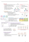
= 20 principle amino acids found in proteins, but other non- Consists of;
protein amino acids found in all organisms. → A chiral -carbon
= At neutral pH exists as dipolar ions. “zwitterions”. → An amine group (NH2/NH3+)
= Amino group is protonated, carboxyl group is de- → A carboxyl group (COOH/COO-
protonated. )
= The -carbon in amino acids is chiral. → Hydrogen atom
= Amino acids can exist in two forms that are → R group
mirror images of each other (“optical active
pairs”), called L and D isomers.
L- and D-Amino Acids:
They have identical properties (colour,
solubility, melting point) under many conditions, but;
→ Different shape alters biological activity.
→ D-isomer can’t be digested.
→ Can’t interact with tRNA (steric hinderance).
→ Can be found in some proteins in both pro and eukaryotes
(antibiotics, hormones, neuropeptides).
R Groups of Amino Acids
o Varies between amino acids. 20 different R groups
define the 20 amino acids found in proteins.
o Chemical nature of R group determines the
properties of the amino acid.
o Features of R groups; size, shape, charge,
hydrogen bonding capacity, hydrophobicity, chemical reactivity.
Glycine→ smallest amino acid. R Glycine, Alanine, Valine, Leucine, Isoleucine & Methionine
group is a hydrogen atom. → All have aliphatic R groups of increasing size.
→ The longer the aliphatic chain the more hydrophobic the amino acid.
→ Hydrophobic amino acids are often found on the inside of proteins away
from the aqueous cellular environment.
→ Methionine contains a thioether
(-S-) group which contains a sulphur
atom.
Proline→ Has cyclic aliphatic R group.
Unique in that R group is bonded to amino
group and -carbon. More structurally
restricted. Often found in bends in proteins.
Phenylalanine, Tyrosine & Tryptophan
→ Called aromatic amino acids.
→ All contain a phenyl ring.
→ All hydrophobic.
→ Tyrosine and Tryptophan have some hydrophilic properties due to
-OH and -NH groups respectively.
→ -OH group in Tyrosine quite reactive.
1
,Serine & Threonine
→ Similar to Alanine and Valine.
→ -OH groups makes them hydrophilic (usually found on outside of protein).
→ -OH also very reactive.
Cysteine→ similar to Serine. Contains reactive thiol group (-SH).
Pairs of third groups come together to form disulphide bonds.
Lysine & Arginine
→ Basic amino acids.
→ Contain side chains that are positively charged at neutral pH.
Histidine→ basic amino acid. Contains imidazole ring. Has pKa of
6 so can be charged or uncharged or uncharged at pH near to
neutral. Often found at the active site of enzymes where it
can bind and release protons. It is an important buffer of pH
in blood.
Aspartic Acid & Glutamic Acid
→ Acidic amino acid.
→ Sometimes called Aspartate and Glutamate.
→ Negatively charged at physiological pH.
→ Have carboxyl groups (-COOH) at end of side chain.
Asparagine & Glutamine
→ Uncharged derivatives of Aspartate and Glutamate.
→ NH2 group replaces O- in carboxyl group.
Biological Functions of Proteins
- Structural (collagen, keratin)
- Movement (actin, myosin)
- Enzymes and catalysts (trypsin, DNA pol)
- Transport (haemoglobin, transferrin)
- Membrane transport (Na+/K+ pump)
- Hormones (insulin)
- Receptor (acetylcholine receptor
- Defence (antibodies, clotting factor\0
- Chromosome sorting (tubulin)
Molecular Measurements
Unit of mass is the Dalton (Da).
One Da is equivalent to the molecular wight of Hydrogen atom.
Mass of proteins often quoted in kDa (1kDa=1000Da)
Unit of distance is the Angstrom (Å)
1 Å= 1-10m= 0.1nm
2
, Protein Structures
1. Primary: sequence of amino acid in peptide chain (many disulphide
linkages).
2. Secondary: folding/coiling of peptide chain (usually into a -helix or -
pleated sheet)
3. Tertiary: peptide chain folds upon itself.
4. Quaternary: folded peptide chains, join together.
*Polypeptides have direction (polarity). Order of residues is read from the
amino terminal to the carboxy ( C) terminal. The order of amino acid
residues in a protein is called its sequence. Each protein has its own
unique sequence.
Protein Assembly
• Hydrogen removed from amino group.
• Oxygen (or hydroxide) group from
carboxyl group.
• Water released (condensation reaction).
• Resulting covalent bond is peptide bond
Peptide Bond
- Has double bond characteristics; rigid bond, prevents rotation, distance between CO and NH groups
shortened.
- Bond is planar (i.e. in 2 peptides 6 atoms lie in the same plane).
- Bond can exist in trans or cis form. In protein bond is almost always in
trans form (H of amino group trans to O of carboxy group).
- Cis peptide bonds only seen where bond is between proline and any
other amino acid.
- X-pro has steric problems in both trans and cis configurations so both
possible.
The amino acids in a polypeptide are called
residues.
Polypeptide has regular repeating part called the
main chain which is referred to as the backbone.
Size of Peptides and Proteins
• Dipeptides: a few naturally occurring examples aspartame (Asp-Phe)-
artificial sweetener.
• Tripeptides: glutathione (Glu-Cys-Gly)- natural antioxidant.
• Short Polypeptides (10-40aa): peptide hormones. E.g glucagon (29aa).
Neurotransmitters e.g. substance P (10aa).
• Large polypeptides (proteins) (40aa): dystrophin (3684aa)
Protein Confirmation
→ The 3D arrangement of protein atoms in its structure.
→ Confirmation is independent of the number of chains in a protein.
→ Proteins must be in the correct 3-dimentional confirmation to work. They
are translated as linear arrays.
→ Native structure of a protein is the three-dimensional structure of a
protein under physiological conditions.
3
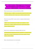
, Anfinsen’s Experiments Ribonuclease A
He performed some experiments investigating protein folding. - Hydrolyses RNA
He used solutions containing 8M urea or 6M guanidine hydrochloride. In - 124 amino acids (Bovine)
these solutions proteins denature (unfold) into random coils. - 4 disulphide links in native
Also used -mercaptoethanol which is a reducing agent and breaks conformation: 26-84,40-95,
disulphide bonds. 58-110, 65-72
Anfinsen dissolved RNAse in solution of -mercaptoethanol and 8M urea.
RNAse losses all activity (completely denatured).
He concluded that the amino acid sequence of RNAse provides all the
information required to specify its native structure.
Conclusion: the primary structure (the amino acid sequence) contains all the information to correctly fold
the protein into its final conformation.
Further Experiment: Anfinsen denatured RNAse in urea and -
mercaptoethanol. Then removed the -mercaptoethanol but left the urea.
Observed that RNAse regained 1% of original activity. This is because there
is only 1 combination of correct disulphide bond but 105 possible
combinations. In urea disulphide bonds form randomly in 1 out of 105
possible combinations the correct bond pattern is obtained 1/105=1%. He added back trace amounts of -
mercaptoethanol to scrambled RNAse (104 incorrect forms of RNAse). After 10 hours the RNAse has
completely regained is activity. Anfinsen’s explanation was that the correct disulphide bonds were in a
lower free energy state and are more energetically favourable.
The thermodynamically most stable structure of RNAse is its native conformation. This I true for all
proteins.
Levinthal Paradox:
- In 1969 Cyrus Levinthal calculated that a protein containing 100 residues would take 1.6x10 27 years to fold
into correct conformation using random chance. But all proteins fold much quicker than this (e.g. RNAse
folds in seconds).
- Levinthal concluded that proteins do not fold via random (stochastic) pathways.
Protein Folding→ proteins fold to their native structures via the formation of partially correct secondary
structures. This model suggests that folding proceeds via a series of intermediates.
Protein Conformation
▪ Proteins are translated as linear arrays.
▪ Proteins must fold into their native conformation.
▪ The principal factor governing folding is that hydrophobic side chains
are buried in the core of the protein while hydrophilic side chains are
exposed on the outside. The exception is integral membrane proteins
where the opposite is true.
Forces that Stabilise Protein Structure
= Interactions between residues (primary structure).
= Interaction of backbones (secondary structure).
= Covalent-disulphide bridges (not all proteins have them).
= Non-covalent-hydrogen bonds, electrostatic
interactions, Van der Waals forces and hydrophobic
Denaturation is the loss of function due to loss of
effect. 3D shape; Heat, pH, Metals, Chemicals, Other
proteins.
Chaperone of Proteins
Cell is not the ideal environment for protein folding.
Busy cell environment.
Proteins not translated instantaneously.
Chaperone proteins help proteins fold correctly.
Delay folding, change environment.
Secondary Structure of a Protein
4
= 20 principle amino acids found in proteins, but other non- Consists of;
protein amino acids found in all organisms. → A chiral -carbon
= At neutral pH exists as dipolar ions. “zwitterions”. → An amine group (NH2/NH3+)
= Amino group is protonated, carboxyl group is de- → A carboxyl group (COOH/COO-
protonated. )
= The -carbon in amino acids is chiral. → Hydrogen atom
= Amino acids can exist in two forms that are → R group
mirror images of each other (“optical active
pairs”), called L and D isomers.
L- and D-Amino Acids:
They have identical properties (colour,
solubility, melting point) under many conditions, but;
→ Different shape alters biological activity.
→ D-isomer can’t be digested.
→ Can’t interact with tRNA (steric hinderance).
→ Can be found in some proteins in both pro and eukaryotes
(antibiotics, hormones, neuropeptides).
R Groups of Amino Acids
o Varies between amino acids. 20 different R groups
define the 20 amino acids found in proteins.
o Chemical nature of R group determines the
properties of the amino acid.
o Features of R groups; size, shape, charge,
hydrogen bonding capacity, hydrophobicity, chemical reactivity.
Glycine→ smallest amino acid. R Glycine, Alanine, Valine, Leucine, Isoleucine & Methionine
group is a hydrogen atom. → All have aliphatic R groups of increasing size.
→ The longer the aliphatic chain the more hydrophobic the amino acid.
→ Hydrophobic amino acids are often found on the inside of proteins away
from the aqueous cellular environment.
→ Methionine contains a thioether
(-S-) group which contains a sulphur
atom.
Proline→ Has cyclic aliphatic R group.
Unique in that R group is bonded to amino
group and -carbon. More structurally
restricted. Often found in bends in proteins.
Phenylalanine, Tyrosine & Tryptophan
→ Called aromatic amino acids.
→ All contain a phenyl ring.
→ All hydrophobic.
→ Tyrosine and Tryptophan have some hydrophilic properties due to
-OH and -NH groups respectively.
→ -OH group in Tyrosine quite reactive.
1
,Serine & Threonine
→ Similar to Alanine and Valine.
→ -OH groups makes them hydrophilic (usually found on outside of protein).
→ -OH also very reactive.
Cysteine→ similar to Serine. Contains reactive thiol group (-SH).
Pairs of third groups come together to form disulphide bonds.
Lysine & Arginine
→ Basic amino acids.
→ Contain side chains that are positively charged at neutral pH.
Histidine→ basic amino acid. Contains imidazole ring. Has pKa of
6 so can be charged or uncharged or uncharged at pH near to
neutral. Often found at the active site of enzymes where it
can bind and release protons. It is an important buffer of pH
in blood.
Aspartic Acid & Glutamic Acid
→ Acidic amino acid.
→ Sometimes called Aspartate and Glutamate.
→ Negatively charged at physiological pH.
→ Have carboxyl groups (-COOH) at end of side chain.
Asparagine & Glutamine
→ Uncharged derivatives of Aspartate and Glutamate.
→ NH2 group replaces O- in carboxyl group.
Biological Functions of Proteins
- Structural (collagen, keratin)
- Movement (actin, myosin)
- Enzymes and catalysts (trypsin, DNA pol)
- Transport (haemoglobin, transferrin)
- Membrane transport (Na+/K+ pump)
- Hormones (insulin)
- Receptor (acetylcholine receptor
- Defence (antibodies, clotting factor\0
- Chromosome sorting (tubulin)
Molecular Measurements
Unit of mass is the Dalton (Da).
One Da is equivalent to the molecular wight of Hydrogen atom.
Mass of proteins often quoted in kDa (1kDa=1000Da)
Unit of distance is the Angstrom (Å)
1 Å= 1-10m= 0.1nm
2
, Protein Structures
1. Primary: sequence of amino acid in peptide chain (many disulphide
linkages).
2. Secondary: folding/coiling of peptide chain (usually into a -helix or -
pleated sheet)
3. Tertiary: peptide chain folds upon itself.
4. Quaternary: folded peptide chains, join together.
*Polypeptides have direction (polarity). Order of residues is read from the
amino terminal to the carboxy ( C) terminal. The order of amino acid
residues in a protein is called its sequence. Each protein has its own
unique sequence.
Protein Assembly
• Hydrogen removed from amino group.
• Oxygen (or hydroxide) group from
carboxyl group.
• Water released (condensation reaction).
• Resulting covalent bond is peptide bond
Peptide Bond
- Has double bond characteristics; rigid bond, prevents rotation, distance between CO and NH groups
shortened.
- Bond is planar (i.e. in 2 peptides 6 atoms lie in the same plane).
- Bond can exist in trans or cis form. In protein bond is almost always in
trans form (H of amino group trans to O of carboxy group).
- Cis peptide bonds only seen where bond is between proline and any
other amino acid.
- X-pro has steric problems in both trans and cis configurations so both
possible.
The amino acids in a polypeptide are called
residues.
Polypeptide has regular repeating part called the
main chain which is referred to as the backbone.
Size of Peptides and Proteins
• Dipeptides: a few naturally occurring examples aspartame (Asp-Phe)-
artificial sweetener.
• Tripeptides: glutathione (Glu-Cys-Gly)- natural antioxidant.
• Short Polypeptides (10-40aa): peptide hormones. E.g glucagon (29aa).
Neurotransmitters e.g. substance P (10aa).
• Large polypeptides (proteins) (40aa): dystrophin (3684aa)
Protein Confirmation
→ The 3D arrangement of protein atoms in its structure.
→ Confirmation is independent of the number of chains in a protein.
→ Proteins must be in the correct 3-dimentional confirmation to work. They
are translated as linear arrays.
→ Native structure of a protein is the three-dimensional structure of a
protein under physiological conditions.
3
, Anfinsen’s Experiments Ribonuclease A
He performed some experiments investigating protein folding. - Hydrolyses RNA
He used solutions containing 8M urea or 6M guanidine hydrochloride. In - 124 amino acids (Bovine)
these solutions proteins denature (unfold) into random coils. - 4 disulphide links in native
Also used -mercaptoethanol which is a reducing agent and breaks conformation: 26-84,40-95,
disulphide bonds. 58-110, 65-72
Anfinsen dissolved RNAse in solution of -mercaptoethanol and 8M urea.
RNAse losses all activity (completely denatured).
He concluded that the amino acid sequence of RNAse provides all the
information required to specify its native structure.
Conclusion: the primary structure (the amino acid sequence) contains all the information to correctly fold
the protein into its final conformation.
Further Experiment: Anfinsen denatured RNAse in urea and -
mercaptoethanol. Then removed the -mercaptoethanol but left the urea.
Observed that RNAse regained 1% of original activity. This is because there
is only 1 combination of correct disulphide bond but 105 possible
combinations. In urea disulphide bonds form randomly in 1 out of 105
possible combinations the correct bond pattern is obtained 1/105=1%. He added back trace amounts of -
mercaptoethanol to scrambled RNAse (104 incorrect forms of RNAse). After 10 hours the RNAse has
completely regained is activity. Anfinsen’s explanation was that the correct disulphide bonds were in a
lower free energy state and are more energetically favourable.
The thermodynamically most stable structure of RNAse is its native conformation. This I true for all
proteins.
Levinthal Paradox:
- In 1969 Cyrus Levinthal calculated that a protein containing 100 residues would take 1.6x10 27 years to fold
into correct conformation using random chance. But all proteins fold much quicker than this (e.g. RNAse
folds in seconds).
- Levinthal concluded that proteins do not fold via random (stochastic) pathways.
Protein Folding→ proteins fold to their native structures via the formation of partially correct secondary
structures. This model suggests that folding proceeds via a series of intermediates.
Protein Conformation
▪ Proteins are translated as linear arrays.
▪ Proteins must fold into their native conformation.
▪ The principal factor governing folding is that hydrophobic side chains
are buried in the core of the protein while hydrophilic side chains are
exposed on the outside. The exception is integral membrane proteins
where the opposite is true.
Forces that Stabilise Protein Structure
= Interactions between residues (primary structure).
= Interaction of backbones (secondary structure).
= Covalent-disulphide bridges (not all proteins have them).
= Non-covalent-hydrogen bonds, electrostatic
interactions, Van der Waals forces and hydrophobic
Denaturation is the loss of function due to loss of
effect. 3D shape; Heat, pH, Metals, Chemicals, Other
proteins.
Chaperone of Proteins
Cell is not the ideal environment for protein folding.
Busy cell environment.
Proteins not translated instantaneously.
Chaperone proteins help proteins fold correctly.
Delay folding, change environment.
Secondary Structure of a Protein
4