MSK 2 – Knie – Isabel Baert
Inleiding – Articulatio genu
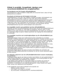
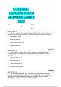
Samengesteld gewricht: tibiofemoraal + patellofemoraal gewricht
1. Tibiofemoraal gewricht
Gevormd door de condyli femores en condyli tibiae
Gewrichtsvlakken
Femurcondylen = convex
• Verschillend in grootte
• Verschillend in kromming (mediale condylus is sterker gekromd)
ð Zorgen voor optredende rotatie tijdens flexie en extensie
ð Volledige extensie = slotrotatie
o Exo van tibia tov femur of endo van femur tov tibia
Tibiaplateau en menisci = concaaf
• Discongruente verbinding tussen de femurcondylen en het tibiaplateau
• Dikke kraakbeenlaag vangt dit gedeeltelijk op, ook menisci zorgen voor betere congruentie
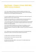
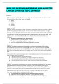
Menisci
Mediale en laterale meniscus, elk voorhoorn en achterhoorn
Functies menisci
• Opheffen discongruentie met toename in stabiliteit
• Gelijkmatigere drukverdeling
• Verdeling van het synoviale vocht
• Schokabsorptie
• Faciliteren van het fysiologisch artrokinematisch bewegingsgedrag
Functionele verbindingen menisci
Verbindingen …
• Met gewrichtskapsel
• Met tibia (meniscotibiale lig)
• Met femur (meniscofemorale lig)
• Met patella
• Tussen mediale meniscus en lig coll mediale
• Tussen mediale/laterale meniscus en strekapparaat
• Tussen laterale meniscus en tractus iliotibialis, m popliteus en m biceps femoris
, • Tussen mediale meniscus en m semimembranosus
• Tussen voorhoorn mediale meniscus en voorhoorn laterale meniscus (lig trans genus)
Mediale meniscus
Veel hechter verbonden dan laterale à laterale een grotere beweeglijkheid
ð Mediale meniscus is kwetsbaarder bij extreme gewrichtsbewegingen
Menisci bewegingen
• Bij flexie en extensie beweging volgen de menisci de tibia
• Bij rotaties volgen de menisci de femurcondylen
Buitenste derde van de menisci
• Bloedvoorziening (vooral voorste en achterste hoorn)
• Rest van de voeding gebeurt via synoviale vloeistoffen
Stabilisatoren: passief (kapselband) & actief (musculatuur)
Door discongruentie is er enkel stabiliteit in de eindposities (flexie en extensiestand)
• Iedere tussenpositie is een atrogeen instabiele stand à krijgen slechts stabiliteit door /p/ en
/a/ stabilisatoren (hoge eisen gesteld aan deze structuren)
Gewrichtsbanden zijn verdikkingen van gewrichtskapsel, uitgezonderd het extracapsulair gelegen lig
coll mediale
1. Mediale compartiment
Passief
• Anterieur deel
o Meniscofemorale ligamenten
o Meniscotibiale ligamenten
• Intermediair deel
o Meniscofemorale ligamenten
o Meniscotibiale ligamenten
o Ligamentum collaterale mediale
• Posterieur deel
o Meniscofemorale ligamenten
o Meniscotibiale ligamenten
o Ligamentum posterior obliquum
o Ligamentum popliteum obliquum
Actief
• Anterieur deel
o Retinaculum extensorum
o M vastus medialis
, • Posterieur deel
o M gastrocnemius
o M semitendinosus
o M semimembranosus
2. Laterale compartiment
Passief
• Anterieur deel
o Meniscofemorale ligamenten
o Meniscotibiale ligamenten
• Intermediair deel
o Meniscofemorale ligamenten
o Meniscotibiale ligamenten
o Ligamentum iliotibiale
• Posterieur deel
o Meniscofemorale ligamenten
o Meniscotibiale ligamenten
o Ligamentum popliteum arcuatum
o Ligamentum collaterale laterale (extra – kapsulair)
Actief
• Anterieur deel
o Retinaculum extensorum
o M vastus lateralis
• Intermediair deel
o Tractus iliotibialis
• Posterieur deel
o M gastrocnemius
o M biceps femoris
o M popliteus
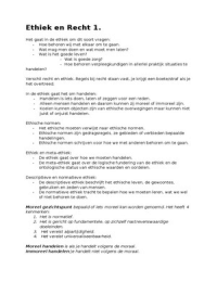
, 3. Centrale compartiment
Passief
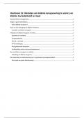
• Voorste kruisband
o Verloop: van centraal anterieur deel van tibia naar achterste binnenste deel van
laterale condylus van femur (craniaal lateraal verloop
o Functie: beperkt de schuifbeweging van tibia naar ventraal tov de femur
o Bij hyperextensie is de VKB maximaal gespannen (ook AKB komt onder een zekere
spanning)
• Achterste kruisband
o Verloop: loopt van centrale achterzijde van tibia naar het midden van binnenzijde van
de mediale condylus van de femur (craniaal mediaal verloop)
o Functie: beperkt de schuifbeweging van tibia naar dorsaal tov femur
o Bij maximale flexie staat vooral het voorste deel van AKB onder spanning
Opmerking
• Exorotatie: kruisbanden ontspannen
• Endorotatie: kruisbanden worden opgespannen (draaien in elkaar)
• Het op spanning komen van de VKB op het einde van extensie zorgt mee voor de exo van de
tibia, slotexorotatie
• Mediale compartiment + centrale compartiment = mediale complex
• Laterale compartiment + centrale compartiment = laterale complex
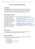
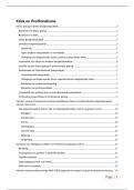
2. Patellofemoraal gewricht
Gevormd door facies articularis patellae van de patella en de facies patellaris van het femur
Patella à beweeglijk verbonden door een kruisvormig weke – delen systeem
• Passieve en actieve elementen die bewegingen van patella geleiden en stabiliseren
Passieve elementen
Centraal
• Lig patellae
Lateraal
• Lig patellofemorale laterale
• Lig meniscopatellare laterale
• Laterale en patellaire expansie van de fascia latae
Mediaal
• Lig patellofemorale mediale
• Lig meniscopatellare mediale
Inleiding – Articulatio genu
Samengesteld gewricht: tibiofemoraal + patellofemoraal gewricht
1. Tibiofemoraal gewricht
Gevormd door de condyli femores en condyli tibiae
Gewrichtsvlakken
Femurcondylen = convex
• Verschillend in grootte
• Verschillend in kromming (mediale condylus is sterker gekromd)
ð Zorgen voor optredende rotatie tijdens flexie en extensie
ð Volledige extensie = slotrotatie
o Exo van tibia tov femur of endo van femur tov tibia
Tibiaplateau en menisci = concaaf
• Discongruente verbinding tussen de femurcondylen en het tibiaplateau
• Dikke kraakbeenlaag vangt dit gedeeltelijk op, ook menisci zorgen voor betere congruentie
Menisci
Mediale en laterale meniscus, elk voorhoorn en achterhoorn
Functies menisci
• Opheffen discongruentie met toename in stabiliteit
• Gelijkmatigere drukverdeling
• Verdeling van het synoviale vocht
• Schokabsorptie
• Faciliteren van het fysiologisch artrokinematisch bewegingsgedrag
Functionele verbindingen menisci
Verbindingen …
• Met gewrichtskapsel
• Met tibia (meniscotibiale lig)
• Met femur (meniscofemorale lig)
• Met patella
• Tussen mediale meniscus en lig coll mediale
• Tussen mediale/laterale meniscus en strekapparaat
• Tussen laterale meniscus en tractus iliotibialis, m popliteus en m biceps femoris
, • Tussen mediale meniscus en m semimembranosus
• Tussen voorhoorn mediale meniscus en voorhoorn laterale meniscus (lig trans genus)
Mediale meniscus
Veel hechter verbonden dan laterale à laterale een grotere beweeglijkheid
ð Mediale meniscus is kwetsbaarder bij extreme gewrichtsbewegingen
Menisci bewegingen
• Bij flexie en extensie beweging volgen de menisci de tibia
• Bij rotaties volgen de menisci de femurcondylen
Buitenste derde van de menisci
• Bloedvoorziening (vooral voorste en achterste hoorn)
• Rest van de voeding gebeurt via synoviale vloeistoffen
Stabilisatoren: passief (kapselband) & actief (musculatuur)
Door discongruentie is er enkel stabiliteit in de eindposities (flexie en extensiestand)
• Iedere tussenpositie is een atrogeen instabiele stand à krijgen slechts stabiliteit door /p/ en
/a/ stabilisatoren (hoge eisen gesteld aan deze structuren)
Gewrichtsbanden zijn verdikkingen van gewrichtskapsel, uitgezonderd het extracapsulair gelegen lig
coll mediale
1. Mediale compartiment
Passief
• Anterieur deel
o Meniscofemorale ligamenten
o Meniscotibiale ligamenten
• Intermediair deel
o Meniscofemorale ligamenten
o Meniscotibiale ligamenten
o Ligamentum collaterale mediale
• Posterieur deel
o Meniscofemorale ligamenten
o Meniscotibiale ligamenten
o Ligamentum posterior obliquum
o Ligamentum popliteum obliquum
Actief
• Anterieur deel
o Retinaculum extensorum
o M vastus medialis
, • Posterieur deel
o M gastrocnemius
o M semitendinosus
o M semimembranosus
2. Laterale compartiment
Passief
• Anterieur deel
o Meniscofemorale ligamenten
o Meniscotibiale ligamenten
• Intermediair deel
o Meniscofemorale ligamenten
o Meniscotibiale ligamenten
o Ligamentum iliotibiale
• Posterieur deel
o Meniscofemorale ligamenten
o Meniscotibiale ligamenten
o Ligamentum popliteum arcuatum
o Ligamentum collaterale laterale (extra – kapsulair)
Actief
• Anterieur deel
o Retinaculum extensorum
o M vastus lateralis
• Intermediair deel
o Tractus iliotibialis
• Posterieur deel
o M gastrocnemius
o M biceps femoris
o M popliteus
, 3. Centrale compartiment
Passief
• Voorste kruisband
o Verloop: van centraal anterieur deel van tibia naar achterste binnenste deel van
laterale condylus van femur (craniaal lateraal verloop
o Functie: beperkt de schuifbeweging van tibia naar ventraal tov de femur
o Bij hyperextensie is de VKB maximaal gespannen (ook AKB komt onder een zekere
spanning)
• Achterste kruisband
o Verloop: loopt van centrale achterzijde van tibia naar het midden van binnenzijde van
de mediale condylus van de femur (craniaal mediaal verloop)
o Functie: beperkt de schuifbeweging van tibia naar dorsaal tov femur
o Bij maximale flexie staat vooral het voorste deel van AKB onder spanning
Opmerking
• Exorotatie: kruisbanden ontspannen
• Endorotatie: kruisbanden worden opgespannen (draaien in elkaar)
• Het op spanning komen van de VKB op het einde van extensie zorgt mee voor de exo van de
tibia, slotexorotatie
• Mediale compartiment + centrale compartiment = mediale complex
• Laterale compartiment + centrale compartiment = laterale complex
2. Patellofemoraal gewricht
Gevormd door facies articularis patellae van de patella en de facies patellaris van het femur
Patella à beweeglijk verbonden door een kruisvormig weke – delen systeem
• Passieve en actieve elementen die bewegingen van patella geleiden en stabiliseren
Passieve elementen
Centraal
• Lig patellae
Lateraal
• Lig patellofemorale laterale
• Lig meniscopatellare laterale
• Laterale en patellaire expansie van de fascia latae
Mediaal
• Lig patellofemorale mediale
• Lig meniscopatellare mediale