Brain Imaging Techniques – MRI, CT, PET & fMRI Explained _ Study Guide Page 1 2026-03-18
Brain Imaging Techniques –
MRI, CT, PET & fMRI
Explained | Study Guide
Page 1 of 6Guidehttps://www.stuvia.com/dashboard!@_)#*)(@$)($@*($@)($@*_ 1 Brain Imaging Techniques – MRI, CT, PET & fMRI Explained _ Study Guide.pdf
, Brain Imaging Techniques Page 2 2026-03-18
MEG
A technique that can quantitatively measure the strength of activity in various regions of the brain. Reveals source of
weak magnetic fields emitted by neurons. Cylinder shaped sensors by head monitor magnetic field. Unlike other
techniques, it can characterize rapidly changes of neural activity at a HIGH RESolution. Also shows how long neural
activation is sustained in brain. Scientists use info fromfMRI and MEG because fMRI provides info about areas of brain
activity in a specific task while mEG tells when certain areas become active (qualitative)
fMRI
detecting changes in oxygen content of the blood (hemoglobin) as it responds to input reaching each brain area
MRI
Provides a high quality, three dimensional image of organs and structure inside body. Noninvasive and detailed. Tell
scientists about structural abnormalities when they first appear, and their progression. Can also reveal very small
changes.
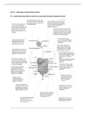
It requires a 15 minute procedure. A patient lies in a hollow tube. A background magnetic field that all atoms in the
brain resonate to is turned on. A second magnetic field is turned that only some atoms resonate too. The second
magnetic field is turned on and of several times, and the atoms creeate an image. Tissue that contains a lot of fat and
water makes a bright image, and tissue that contains little or no water b/c water reacts to the magnetic field. (bone)
would create a black image. Valuable for studying brain and spinal cord. We can see tumors and early damage from
stroke.
Page 2 of 6 2 Brain Imaging Techniques.pdf
Brain Imaging Techniques –
MRI, CT, PET & fMRI
Explained | Study Guide
Page 1 of 6Guidehttps://www.stuvia.com/dashboard!@_)#*)(@$)($@*($@)($@*_ 1 Brain Imaging Techniques – MRI, CT, PET & fMRI Explained _ Study Guide.pdf
, Brain Imaging Techniques Page 2 2026-03-18
MEG
A technique that can quantitatively measure the strength of activity in various regions of the brain. Reveals source of
weak magnetic fields emitted by neurons. Cylinder shaped sensors by head monitor magnetic field. Unlike other
techniques, it can characterize rapidly changes of neural activity at a HIGH RESolution. Also shows how long neural
activation is sustained in brain. Scientists use info fromfMRI and MEG because fMRI provides info about areas of brain
activity in a specific task while mEG tells when certain areas become active (qualitative)
fMRI
detecting changes in oxygen content of the blood (hemoglobin) as it responds to input reaching each brain area
MRI
Provides a high quality, three dimensional image of organs and structure inside body. Noninvasive and detailed. Tell
scientists about structural abnormalities when they first appear, and their progression. Can also reveal very small
changes.
It requires a 15 minute procedure. A patient lies in a hollow tube. A background magnetic field that all atoms in the
brain resonate to is turned on. A second magnetic field is turned that only some atoms resonate too. The second
magnetic field is turned on and of several times, and the atoms creeate an image. Tissue that contains a lot of fat and
water makes a bright image, and tissue that contains little or no water b/c water reacts to the magnetic field. (bone)
would create a black image. Valuable for studying brain and spinal cord. We can see tumors and early damage from
stroke.
Page 2 of 6 2 Brain Imaging Techniques.pdf