Anatomie dissecties - practicumvoorbereiding KV BA 2 SEM 2
Anatomie dissecties - Hoofd & hals (reeks A)
A1: Hals oppervlakkig (ventraal en lateraal) _____________________________________________ 1
A3: Trigonum caroticum ____________________________________________________________ 5
A4: Regio colli mediana _____________________________________________________________ 8
A5: Aangezicht - Regio parotideamaseterica en parotisloge _______________________________ 10
A6: Kauwspieren - fossa temporalis - fossa infratemporalis _______________________________ 15
A8: De schedelholte ______________________________________________________________ 24
A9: De orbita ____________________________________________________________________ 27
A10: Pharynx - larynx - tong ________________________________________________________ 33
A1: Hals oppervlakkig (ventraal en lateraal)
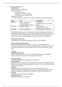
platysma: in oppervlakkige cervicale fascia
⤷ tussen huid en diepe cervicale fascia
origo: Huid / fascia van infra- en supraclaviculaire regio's
insertie: Lower border of mandible, skin of buccal/cheek region, lower lip, modiolus, orbicularis
oris muscle
n. transversus colli
van: cervical plexus (C2 and C3)
verloop:
- draait rond rand sternocleidomastoideus
- gaat daar horizontaal over
- gaat onder v. jugularis externa
- perforeert diepe cervicale fascia
- splitst in: stijgende en dalende takken (onder platysma)
v. jugularis anterior
draineert in: v. jugularis extarna (occasioneel in v. subclavia)
ligt lateraal v cricothyroid ligament
verloop:
- daalt af tssn mediane lijn en voorste rand vd sternocleidomastoideus,
- onderste deel nek: gaat onder sternocleidomastoideus
- draineert dan in v. jugularis externa
1
,Anatomie dissecties - practicumvoorbereiding KV BA 2 SEM 2
Net boven borstbeen: de 2 venen met elkaar in verbinding via arcus venosus
jugularis
⤷ ontvangt ook zijrivieren vd vv. thyroidea inferiores (die ook communiceren met
v. jug interna)
(cutane) takken van de plexus cervicalis
N. occipitalis minor
ontstaan: komt voort uit laterale tak vd ventrale ramus vd 2de cervicale zenuw
verloop: buigt rond en stijgt langs achterste rand van de Sternocleidomastoideus richting schedel
N. auricularis magnus
→ sensorische innervatie v huid over glandula parotis/parotidea, processus mastoideus en opp v
uitw oor
grootste vd stijgende takken vd cervicale plexus
ontstaat uit: 2de en 3de cervicale zenuw
verloop:
- rond posterieure rand sternocleidomastoideus
- door diepe fascia
- stijgt onder platysma nr glandula parotidea
⤷ splitst daar in anterieure en post tak
N. transversus colli
zie boven
Nn. supraclaviculares mediales, intermedii en laterales
→ innerveren huid vd schouder
ontstaan: C3 - C4
verloop:
- komen tevoorschijn onder achterste rand sternocleidomastoideus
- dalen af in achterste halsdriehoek onder platysma en diepe cervicale fascia
mediale: kruist schuin over v. jugularis ext en claviculaire en sternale kop v sternocleidomastoideus
intermedii: kruist de clavicula
laterales: gaan schuin over buitenoppervlak van de trapezius en het acromion
fascia cervicalis superficialis en de M. omohyoideus
fascia cervicalis superficialis: dunne laag onderhuids bindweefsel dat tussen dermis vd huid en
investing layer v fascia cervicalis profundus ligt
bevat:
- platysma
- cutane zenuwen, bloed- en lymfevaten, oppervlakkige lymfeklieren
2
,Anatomie dissecties - practicumvoorbereiding KV BA 2 SEM 2
m. omohyoideus
→ aanspannen halsfacia dr vergroeiing v zijn tussenpees met vagina carotica
→ openhouden v. jugularis interna
→ naar caudaal trekken trongbeen
→ fixeren tongbeen
Origo: venter inferior: margo superior scapula, boden proc coracoideus
insertie: venter superior: corpus ossis hyoidei
Craniaal van de M. omohyoideus
plexus cervicalis
→ w gevormd dr anterieure rami v cervicale zenuwen C1 tot C4
anostomose met: n. accessorius, n. hypoglossus en truncus sympaticus
takken v cervicale plexus komen uit posterieure halsdriehoek bij punctum nervosum, punt dat
halverwege op achterste rand v sternocleidomastoideum ligt
Cutaan (4 takken):
- n. occipitalis major - (C2, posterieure ramus)
- n. occipitalis minor - innerveert huid en hoofdhuid posterosuperior vd oorschelp (C2)
- n. auricularis magnus - innerveert huid nabij concha oorschelp (buitenoor) en externe
akoestische gehoorgang (gehoorgang) (C2 & C3)
- n. transversus colli - innerveert het voorste deel van de nek (C2 en C3)
- Supraclaviculaire zenuwen - innerveren huid boven en onder sleutelbeen (C3 en C4)
musculaire takken:
- Ansa cervicalis (lus gevormd uit C1-C3 die de 4 infrahyoid of
‘strapmuscles’ bezenuwt)
- n. phrenicus (C3-C5 (vnl C4)) - innerveren diafragma en
pericard
- Segmentale takken (C1-C4) - innerveren anterieure en
middelste scaleni
3
,Anatomie dissecties - practicumvoorbereiding KV BA 2 SEM 2
Plexus brachialis
Caudaal van de M. omohyoideus
De fossa supraclavicularis major of het trigonum omoclaviculare.
onderverdeeld in fossa supraclavicularis major and fossa supraclavicularis minor
⤷ door: m. sternocleidomastoideus (minor: tssn de 2 koppen vd sternocleidom.)
Achter de fascia colli media of de lamina pretrachealis
lamina pretrachealis
Nodi lymphoidei cervicales laterales
boven n. accessorius
nabij bovenste deel v. jugularis interna, lateraal of posterieur vd vagina carotica
nodi profundi inferiores.
vlak naast v. jugularis interna tussen interna en externa
4
,Anatomie dissecties - practicumvoorbereiding KV BA 2 SEM 2
A3: Trigonum caroticum
Ansa cervicalis
= lus id cervicale plexus
bstt uit:
- radix superior: gevormd uit C1,
- radix inferior: gevormd uit C2 en C3
radix superior:
verloop:
- vezels verlopen initieel met n. hypoglossus
- voor a. carotis interna
- hft aftakkingen nr voor nr de strap muscles
⤷ venter superior vd omohyoideus, geniohyoideus
en thyrohyoideus
radix inferior
verloop: (meer posterolateraal)
- voor v. jugularis interna
- hft takken nr beneden
⤷ venter inferior vd omohyoideus, inferieure delen vd sternohyoideus en sternothyroideus
n. laryngeus superior
tak vd n. vagus
⤷ ontstaat uit ganglion inferior vd n. vagus
⤷ ontvangt tak vh ganglion cervicale inferior
verloop:
- daalt af langs farynx achter a. carotis interna
- splitst in 2 takken: externa en interna (mediaal vd a. carotis interna)
kliniek: verlamming v deze zenuw: verandert toonhoogte vd stem en zrgt vr het niet meer knnn
maken v explosief geluid (dr verlamming cricothyroid spier)
bilateraal: paardachtige en vermoeide stem
Ramus internus n. laryngeus
verloop:
- daalt nr thyrohyoid membraan (fibreus membraan vd larynx onder os hyoideum)
⤷ samen met a. laryngea superior (tak vd a. thyroidea superior)
Ramus externus n. laryngeus
verloop:
- daalt af op larynx, onder m. sternothyroideus
- nr m. cricothyroideus
5
, Anatomie dissecties - practicumvoorbereiding KV BA 2 SEM 2
sinus caroticus
sinus caroticus
= verwijding aan basis a. carotis interna met baroreceptoren die verandering v bloeddruk registeren
a. occiptialis
tak vd a. carotica externa
ontstaan: - rechtegenover a. facialis (nr voor) nr achter
- bedekt dr: venter posterior vd gigastricus en stylohyoideus
verloop:
- kruist a. carotis interna, v. jugularis interna en n. vagus en accesorius
- achter processus mastoideus rochting os occipitale
n. vagus
- uit foramen jugulare
- loopt tssn a. carotis interna en v. jugularis interna
- voor a. subclavia (R) en aortaboog (L)
⤷ gft aftakking: n. laryngeus recurrens
Fascia cervicalis profunda
ligt onder platysma
onderverdeeld in:
- lamina superficialis fasciae cervicalis (meest oppervlakkig): omgeeft hele nek
- lamina pretrachealis: strekt zich mediaal uit voor aa. carotes en vormt mee vagina carotica
⤷ ligt achter spieren die depressie vh hyoid verzorgen
- lamina prevertebralis: strekt zich mediaal uit achter aa. carotes
⤷ rond vertebrea en preverebrale spieren
M. longus colli
Origo: wervellichaam vd 5de halswervel tot 3de borstwervel, procsessi transversie vd 2de - 5de
halswervel
insertie: proccessi transversie vd 5de - 6de halswervel, wervellich vd 2de - 4de halswervel,
tuberculum anterius vd atlas
6
Anatomie dissecties - Hoofd & hals (reeks A)
A1: Hals oppervlakkig (ventraal en lateraal) _____________________________________________ 1
A3: Trigonum caroticum ____________________________________________________________ 5
A4: Regio colli mediana _____________________________________________________________ 8
A5: Aangezicht - Regio parotideamaseterica en parotisloge _______________________________ 10
A6: Kauwspieren - fossa temporalis - fossa infratemporalis _______________________________ 15
A8: De schedelholte ______________________________________________________________ 24
A9: De orbita ____________________________________________________________________ 27
A10: Pharynx - larynx - tong ________________________________________________________ 33
A1: Hals oppervlakkig (ventraal en lateraal)
platysma: in oppervlakkige cervicale fascia
⤷ tussen huid en diepe cervicale fascia
origo: Huid / fascia van infra- en supraclaviculaire regio's
insertie: Lower border of mandible, skin of buccal/cheek region, lower lip, modiolus, orbicularis
oris muscle
n. transversus colli
van: cervical plexus (C2 and C3)
verloop:
- draait rond rand sternocleidomastoideus
- gaat daar horizontaal over
- gaat onder v. jugularis externa
- perforeert diepe cervicale fascia
- splitst in: stijgende en dalende takken (onder platysma)
v. jugularis anterior
draineert in: v. jugularis extarna (occasioneel in v. subclavia)
ligt lateraal v cricothyroid ligament
verloop:
- daalt af tssn mediane lijn en voorste rand vd sternocleidomastoideus,
- onderste deel nek: gaat onder sternocleidomastoideus
- draineert dan in v. jugularis externa
1
,Anatomie dissecties - practicumvoorbereiding KV BA 2 SEM 2
Net boven borstbeen: de 2 venen met elkaar in verbinding via arcus venosus
jugularis
⤷ ontvangt ook zijrivieren vd vv. thyroidea inferiores (die ook communiceren met
v. jug interna)
(cutane) takken van de plexus cervicalis
N. occipitalis minor
ontstaan: komt voort uit laterale tak vd ventrale ramus vd 2de cervicale zenuw
verloop: buigt rond en stijgt langs achterste rand van de Sternocleidomastoideus richting schedel
N. auricularis magnus
→ sensorische innervatie v huid over glandula parotis/parotidea, processus mastoideus en opp v
uitw oor
grootste vd stijgende takken vd cervicale plexus
ontstaat uit: 2de en 3de cervicale zenuw
verloop:
- rond posterieure rand sternocleidomastoideus
- door diepe fascia
- stijgt onder platysma nr glandula parotidea
⤷ splitst daar in anterieure en post tak
N. transversus colli
zie boven
Nn. supraclaviculares mediales, intermedii en laterales
→ innerveren huid vd schouder
ontstaan: C3 - C4
verloop:
- komen tevoorschijn onder achterste rand sternocleidomastoideus
- dalen af in achterste halsdriehoek onder platysma en diepe cervicale fascia
mediale: kruist schuin over v. jugularis ext en claviculaire en sternale kop v sternocleidomastoideus
intermedii: kruist de clavicula
laterales: gaan schuin over buitenoppervlak van de trapezius en het acromion
fascia cervicalis superficialis en de M. omohyoideus
fascia cervicalis superficialis: dunne laag onderhuids bindweefsel dat tussen dermis vd huid en
investing layer v fascia cervicalis profundus ligt
bevat:
- platysma
- cutane zenuwen, bloed- en lymfevaten, oppervlakkige lymfeklieren
2
,Anatomie dissecties - practicumvoorbereiding KV BA 2 SEM 2
m. omohyoideus
→ aanspannen halsfacia dr vergroeiing v zijn tussenpees met vagina carotica
→ openhouden v. jugularis interna
→ naar caudaal trekken trongbeen
→ fixeren tongbeen
Origo: venter inferior: margo superior scapula, boden proc coracoideus
insertie: venter superior: corpus ossis hyoidei
Craniaal van de M. omohyoideus
plexus cervicalis
→ w gevormd dr anterieure rami v cervicale zenuwen C1 tot C4
anostomose met: n. accessorius, n. hypoglossus en truncus sympaticus
takken v cervicale plexus komen uit posterieure halsdriehoek bij punctum nervosum, punt dat
halverwege op achterste rand v sternocleidomastoideum ligt
Cutaan (4 takken):
- n. occipitalis major - (C2, posterieure ramus)
- n. occipitalis minor - innerveert huid en hoofdhuid posterosuperior vd oorschelp (C2)
- n. auricularis magnus - innerveert huid nabij concha oorschelp (buitenoor) en externe
akoestische gehoorgang (gehoorgang) (C2 & C3)
- n. transversus colli - innerveert het voorste deel van de nek (C2 en C3)
- Supraclaviculaire zenuwen - innerveren huid boven en onder sleutelbeen (C3 en C4)
musculaire takken:
- Ansa cervicalis (lus gevormd uit C1-C3 die de 4 infrahyoid of
‘strapmuscles’ bezenuwt)
- n. phrenicus (C3-C5 (vnl C4)) - innerveren diafragma en
pericard
- Segmentale takken (C1-C4) - innerveren anterieure en
middelste scaleni
3
,Anatomie dissecties - practicumvoorbereiding KV BA 2 SEM 2
Plexus brachialis
Caudaal van de M. omohyoideus
De fossa supraclavicularis major of het trigonum omoclaviculare.
onderverdeeld in fossa supraclavicularis major and fossa supraclavicularis minor
⤷ door: m. sternocleidomastoideus (minor: tssn de 2 koppen vd sternocleidom.)
Achter de fascia colli media of de lamina pretrachealis
lamina pretrachealis
Nodi lymphoidei cervicales laterales
boven n. accessorius
nabij bovenste deel v. jugularis interna, lateraal of posterieur vd vagina carotica
nodi profundi inferiores.
vlak naast v. jugularis interna tussen interna en externa
4
,Anatomie dissecties - practicumvoorbereiding KV BA 2 SEM 2
A3: Trigonum caroticum
Ansa cervicalis
= lus id cervicale plexus
bstt uit:
- radix superior: gevormd uit C1,
- radix inferior: gevormd uit C2 en C3
radix superior:
verloop:
- vezels verlopen initieel met n. hypoglossus
- voor a. carotis interna
- hft aftakkingen nr voor nr de strap muscles
⤷ venter superior vd omohyoideus, geniohyoideus
en thyrohyoideus
radix inferior
verloop: (meer posterolateraal)
- voor v. jugularis interna
- hft takken nr beneden
⤷ venter inferior vd omohyoideus, inferieure delen vd sternohyoideus en sternothyroideus
n. laryngeus superior
tak vd n. vagus
⤷ ontstaat uit ganglion inferior vd n. vagus
⤷ ontvangt tak vh ganglion cervicale inferior
verloop:
- daalt af langs farynx achter a. carotis interna
- splitst in 2 takken: externa en interna (mediaal vd a. carotis interna)
kliniek: verlamming v deze zenuw: verandert toonhoogte vd stem en zrgt vr het niet meer knnn
maken v explosief geluid (dr verlamming cricothyroid spier)
bilateraal: paardachtige en vermoeide stem
Ramus internus n. laryngeus
verloop:
- daalt nr thyrohyoid membraan (fibreus membraan vd larynx onder os hyoideum)
⤷ samen met a. laryngea superior (tak vd a. thyroidea superior)
Ramus externus n. laryngeus
verloop:
- daalt af op larynx, onder m. sternothyroideus
- nr m. cricothyroideus
5
, Anatomie dissecties - practicumvoorbereiding KV BA 2 SEM 2
sinus caroticus
sinus caroticus
= verwijding aan basis a. carotis interna met baroreceptoren die verandering v bloeddruk registeren
a. occiptialis
tak vd a. carotica externa
ontstaan: - rechtegenover a. facialis (nr voor) nr achter
- bedekt dr: venter posterior vd gigastricus en stylohyoideus
verloop:
- kruist a. carotis interna, v. jugularis interna en n. vagus en accesorius
- achter processus mastoideus rochting os occipitale
n. vagus
- uit foramen jugulare
- loopt tssn a. carotis interna en v. jugularis interna
- voor a. subclavia (R) en aortaboog (L)
⤷ gft aftakking: n. laryngeus recurrens
Fascia cervicalis profunda
ligt onder platysma
onderverdeeld in:
- lamina superficialis fasciae cervicalis (meest oppervlakkig): omgeeft hele nek
- lamina pretrachealis: strekt zich mediaal uit voor aa. carotes en vormt mee vagina carotica
⤷ ligt achter spieren die depressie vh hyoid verzorgen
- lamina prevertebralis: strekt zich mediaal uit achter aa. carotes
⤷ rond vertebrea en preverebrale spieren
M. longus colli
Origo: wervellichaam vd 5de halswervel tot 3de borstwervel, procsessi transversie vd 2de - 5de
halswervel
insertie: proccessi transversie vd 5de - 6de halswervel, wervellich vd 2de - 4de halswervel,
tuberculum anterius vd atlas
6