Case 7
LG1: Macroanatomy of skeletal muscle
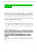
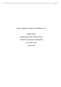
A muscle consists of connective tissue, muscle fibers, nerves & blood vessels.
The connective tissue is divided into three different sheaths:
1. Epimysium (dense irregular tissue that is around the whole muscle);
2. Perimysium and fascicles (muscle fibers are grouped into fascicles which
are surrounded by perimysium)
3. Endomysium (fine areolar connective tissue that wraps around each
individual muscle fiber)
Skeletal muscles are connected to bones and are over wrapping minimum one
joint. They are attached to the connective tissue of the bone either direct to the
bones or indirect via a tendon (aponeurosis).
Shape:
Skeletal muscles vary considerably in size, shape, and arrangement of fibers.
They range from extremely tiny strands such as the stapediusmuscle of the
middle ear to
large masses such as the muscles of the thigh.
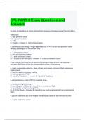
Some skeletal muscles are broad in shape and some narrow. In some muscles
the fibers are:
1. parallel to the long axis of the
muscle;
2. in some they converge to a
narrow attachment;
3. and in some they are oblique.
, LG2:Microanatomy of the muscle
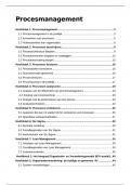
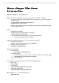
The plasma membrane of the skeletal muscle fiber is called a sarcolemma. The
muscle fiber contains long cylindrical structures, the myofibrils.
The myofibrils almost entirely fill the cell and push the nuclei to the outer edges of
the cell under the sarcolemma. The many myofibrils each have light and dark bands
and are aligned with one another so that the light and dark bands are next to one
another.
This gives the cell its striated appearance
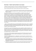
The light bands are called I bands and the dark bands are called A bands. In the
middle of the I band there is a line called the Z line (or disc). In the middle of the A
bands (or dark bands) there is a light zone called the H zone. In the middle of the H
zone there is another line, the M line. The precise arrangement of these features is
due to a chain of functional units in the myofibrils, sarcomeres.
LG1: Macroanatomy of skeletal muscle
A muscle consists of connective tissue, muscle fibers, nerves & blood vessels.
The connective tissue is divided into three different sheaths:
1. Epimysium (dense irregular tissue that is around the whole muscle);
2. Perimysium and fascicles (muscle fibers are grouped into fascicles which
are surrounded by perimysium)
3. Endomysium (fine areolar connective tissue that wraps around each
individual muscle fiber)
Skeletal muscles are connected to bones and are over wrapping minimum one
joint. They are attached to the connective tissue of the bone either direct to the
bones or indirect via a tendon (aponeurosis).
Shape:
Skeletal muscles vary considerably in size, shape, and arrangement of fibers.
They range from extremely tiny strands such as the stapediusmuscle of the
middle ear to
large masses such as the muscles of the thigh.
Some skeletal muscles are broad in shape and some narrow. In some muscles
the fibers are:
1. parallel to the long axis of the
muscle;
2. in some they converge to a
narrow attachment;
3. and in some they are oblique.
, LG2:Microanatomy of the muscle
The plasma membrane of the skeletal muscle fiber is called a sarcolemma. The
muscle fiber contains long cylindrical structures, the myofibrils.
The myofibrils almost entirely fill the cell and push the nuclei to the outer edges of
the cell under the sarcolemma. The many myofibrils each have light and dark bands
and are aligned with one another so that the light and dark bands are next to one
another.
This gives the cell its striated appearance
The light bands are called I bands and the dark bands are called A bands. In the
middle of the I band there is a line called the Z line (or disc). In the middle of the A
bands (or dark bands) there is a light zone called the H zone. In the middle of the H
zone there is another line, the M line. The precise arrangement of these features is
due to a chain of functional units in the myofibrils, sarcomeres.