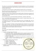
Continuity of Cells
The nucleus of a cell contains all the information for the total functioning of a cell. This information is
carried ono thread-like structures called chromosomes which are made up of DNA and histone proteins,
collectively called chromatin.
In humans there are 46 chromosomes. In people with Downs syndrome, there are 47 chromosomes.
Body cells are said to have a diploid number of chromosomes. These pairs are known as homologous pairs.
Division by mitosis maintains the diploid number.
In gametes the cells are said to be haploid, they have half the number of chromosomes compared to the
diploid number because the gametes only have one chromosome fromo each pair. Haploid cells are
achieved through meiosis.
Each chromosome can carry several thousand pieces of information – each piece is called a gene.
Gene – A short length of DNA that codes for the primary structure of a protein (primary structure – the
sequence of amino acids in the polypeptide chain)
In non-dividing cells, the chromosomes are not visible because they are only partially coiled – they are
more diffusely organised in a form called chromatin. Densely packed is heterochromatin. Less is
euchromatin.
When a cell is preparing for cell division, the DNA replicated and the chromosomes coil up to become
shorter, thicker and denser – this makes then visible under the light microscope as 2 chromatids (sister
chromatids) joined at a specialised area called the centromere. The centromere is the last place to divide.
It holds the whole chromosome together until the final separation.
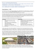
The Cell Cyle
The cell cycle is the complete sequence of events from a cell’s formation until it has divided into two
daughter cells.
Interphase – the longest stage of the cell cycle
o G1 phase (gap or growth) – organelles replicate and other cytoplasmic components replicate.
The cell increases in size. Protein synthesis, RNA synthesis and enzyme synthesis occur.
o S phase (synthesis) – DNA replication takes place (DNA duplication)and chromatids form within
the nucleus. Histones form
o G2 phase – Spindle proteins are synthesised. The chromosomes are checked for errors and
repaired if necessary. A second growth phase takes
place.
Mitosis – the actual division of the chromosomes
o Prophase – preparation stage
o Metaphase – organisation stage
o Anaphase – beginning of separation
o Telophase – completion of separation
Cytokinesis – the division of the cell into two daughter cells
(cytoplasm divides)
, Mitosis
At the end of interphase, DNA has replicated but chromosomes are not yet visible. The nucleus is still
surrounded by its membrane and the nucleolus is still visible.
Prophase
Chromosomes become visible as they condense. During interphase some of the DNA remains unwound
to facilitate the process of protein synthesis. However, when condensed the chromatin has much
greater strength – this is important as it can help to prevent damage during mitosis.
Each chromosome appears as two chromatids,
joined by a centromere.
The nucleolus disappears. In animal cells, centrioles
will move to opposite poles of the cell. Centrioles
are responsible for forming the spindle. The spindle
fibres begin to develop in a star-shape from each
centriole (this star shape is called an aster). In plant
cells, spindle formation takes place without the
presence of centrioles.
By late prophase, the centrioles will have completed
their migration to opposite poles and the spindle
has formed.
The nuclear envelope has broken down.
Metaphase
(meta-middle)
The chromosomes arrange themselves across the equator of the cell. Each
chromosome attaches onto the spindle fibres by their centromere.
Anaphase
The spindle fibres contract, and the centromeres (holding the chromatids of each chromosome
together ) split
Further contraction of the single fibres pulls the chromatids apart.
As anaphase continues, the two chromatids of each chromosome are pulled to
opposite poles of the cell.
The nucleus of a cell contains all the information for the total functioning of a cell. This information is
carried ono thread-like structures called chromosomes which are made up of DNA and histone proteins,
collectively called chromatin.
In humans there are 46 chromosomes. In people with Downs syndrome, there are 47 chromosomes.
Body cells are said to have a diploid number of chromosomes. These pairs are known as homologous pairs.
Division by mitosis maintains the diploid number.
In gametes the cells are said to be haploid, they have half the number of chromosomes compared to the
diploid number because the gametes only have one chromosome fromo each pair. Haploid cells are
achieved through meiosis.
Each chromosome can carry several thousand pieces of information – each piece is called a gene.
Gene – A short length of DNA that codes for the primary structure of a protein (primary structure – the
sequence of amino acids in the polypeptide chain)
In non-dividing cells, the chromosomes are not visible because they are only partially coiled – they are
more diffusely organised in a form called chromatin. Densely packed is heterochromatin. Less is
euchromatin.
When a cell is preparing for cell division, the DNA replicated and the chromosomes coil up to become
shorter, thicker and denser – this makes then visible under the light microscope as 2 chromatids (sister
chromatids) joined at a specialised area called the centromere. The centromere is the last place to divide.
It holds the whole chromosome together until the final separation.
The Cell Cyle
The cell cycle is the complete sequence of events from a cell’s formation until it has divided into two
daughter cells.
Interphase – the longest stage of the cell cycle
o G1 phase (gap or growth) – organelles replicate and other cytoplasmic components replicate.
The cell increases in size. Protein synthesis, RNA synthesis and enzyme synthesis occur.
o S phase (synthesis) – DNA replication takes place (DNA duplication)and chromatids form within
the nucleus. Histones form
o G2 phase – Spindle proteins are synthesised. The chromosomes are checked for errors and
repaired if necessary. A second growth phase takes
place.
Mitosis – the actual division of the chromosomes
o Prophase – preparation stage
o Metaphase – organisation stage
o Anaphase – beginning of separation
o Telophase – completion of separation
Cytokinesis – the division of the cell into two daughter cells
(cytoplasm divides)
, Mitosis
At the end of interphase, DNA has replicated but chromosomes are not yet visible. The nucleus is still
surrounded by its membrane and the nucleolus is still visible.
Prophase
Chromosomes become visible as they condense. During interphase some of the DNA remains unwound
to facilitate the process of protein synthesis. However, when condensed the chromatin has much
greater strength – this is important as it can help to prevent damage during mitosis.
Each chromosome appears as two chromatids,
joined by a centromere.
The nucleolus disappears. In animal cells, centrioles
will move to opposite poles of the cell. Centrioles
are responsible for forming the spindle. The spindle
fibres begin to develop in a star-shape from each
centriole (this star shape is called an aster). In plant
cells, spindle formation takes place without the
presence of centrioles.
By late prophase, the centrioles will have completed
their migration to opposite poles and the spindle
has formed.
The nuclear envelope has broken down.
Metaphase
(meta-middle)
The chromosomes arrange themselves across the equator of the cell. Each
chromosome attaches onto the spindle fibres by their centromere.
Anaphase
The spindle fibres contract, and the centromeres (holding the chromatids of each chromosome
together ) split
Further contraction of the single fibres pulls the chromatids apart.
As anaphase continues, the two chromatids of each chromosome are pulled to
opposite poles of the cell.