

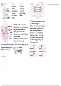
tropomyosin
7
A band
rod cells come cells Iband I band
brain
myosin thick filaments
-
black-white -
Trichromatic - -
IIII
numerous ↳ red , green , blue -
·
t
-
IIII
-
retinal conc & forea
convergencee
-
IIII IIII
-
.
N
for low
light
-
high light intensity ↓
Zline It zone Zline
myosin crossbridge
-
low visual acuity
-
iodopsin Actin thin filaments
-
rhodopSin Sarcomere
+
Ca2 released
T
from sarcoplasmic reticulum
·
·
Ca't binds to tropomyosin
-
Sinoatrial mode generates a wave of excitation
Sinoatrial node
·
change in 30 structure moving tropomyosin
*
-
Wave spreads down the atrial walls (atrial systole
Atrioventricular away from myosin binding site on actin
Activity picked up at atrioventricular node
-
node head (with ADP) binds to act in
myosin
·
- Bundle-slight delay allowing blood into
~ the ventricles
forming actinmyosin cross-bridge
·
of His -wave spread down bundle of His /made up of Purkyne fibres
actin ADP released
angle change pulling
·
L
,
-V to the apex of ventricles
.
Purky ne
r ~ New ATP attaches breaking cross-bridge
↑ V
vv fibers -
wave travels up the walls of the ventricles from the apex
↓
>
·
can t activates ATP
hydrolase moving it back
r
them to contract simultaneously
Tr causing to the original position
·
wave of excitation cannot pass directly to ventricles due to
non-conducting tissue layer -
-
Do ·
ADP
> > ADD
sympathetic/accelerator
Pi
nerve heart rate controlled T
&
Fe
- -
parasympathetic/ragus nerve by medulla
oblongata
,
- -
+
-
& can be stimulated
1
2
by chemical and &
- -
&S
& ~ -
D
r
-
So~ pressure receptors :
·
ADD
ATD L
Pi [ T
- -
- -
7
A band
rod cells come cells Iband I band
brain
myosin thick filaments
-
black-white -
Trichromatic - -
IIII
numerous ↳ red , green , blue -
·
t
-
IIII
-
retinal conc & forea
convergencee
-
IIII IIII
-
.
N
for low
light
-
high light intensity ↓
Zline It zone Zline
myosin crossbridge
-
low visual acuity
-
iodopsin Actin thin filaments
-
rhodopSin Sarcomere
+
Ca2 released
T
from sarcoplasmic reticulum
·
·
Ca't binds to tropomyosin
-
Sinoatrial mode generates a wave of excitation
Sinoatrial node
·
change in 30 structure moving tropomyosin
*
-
Wave spreads down the atrial walls (atrial systole
Atrioventricular away from myosin binding site on actin
Activity picked up at atrioventricular node
-
node head (with ADP) binds to act in
myosin
·
- Bundle-slight delay allowing blood into
~ the ventricles
forming actinmyosin cross-bridge
·
of His -wave spread down bundle of His /made up of Purkyne fibres
actin ADP released
angle change pulling
·
L
,
-V to the apex of ventricles
.
Purky ne
r ~ New ATP attaches breaking cross-bridge
↑ V
vv fibers -
wave travels up the walls of the ventricles from the apex
↓
>
·
can t activates ATP
hydrolase moving it back
r
them to contract simultaneously
Tr causing to the original position
·
wave of excitation cannot pass directly to ventricles due to
non-conducting tissue layer -
-
Do ·
ADP
> > ADD
sympathetic/accelerator
Pi
nerve heart rate controlled T
&
Fe
- -
parasympathetic/ragus nerve by medulla
oblongata
,
- -
+
-
& can be stimulated
1
2
by chemical and &
- -
&S
& ~ -
D
r
-
So~ pressure receptors :
·
ADD
ATD L
Pi [ T
- -
- -


