Biophysics Techniques - X-rays
X-Ray Crystallography
What is it? How does it work?
X-Ray crystallography uses the diffraction of X-Rays in a crystal to determine its structure.
1. An X-ray is generated
• Electrons are generated by heating a tungsten filament, and are then directed toward an
anode, which is kept cool with water and usually made of copper
• The electrons are accelerated through a high voltage (30-40 kV) and undergo rapid
declaration once they hit the target
• They emit Bremsstrahlung (electromagetic) radiation - the energy of the momentum change
is emitted as a photon
2. This is fired at a crystal (biological substance)
• They are diffracted in accordance with Bragg’s Law
• The rays of X-rays are reflected off different layers in the crystal, resulting in a phase
difference
• This phase difference with result in either constructive or destructive interference
• For them to be in phase at the detector, the phase difference must be equal to an integral
number of the wavelength
• n λ = 2d sin θ
3. The pattern of diffraction (interference pattern) is seen on the screen behind it
4. This is used to build an atomic model of the substance
Resolution: atomic
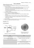
Set Up and Result
Characteristic pattern of a
copper sulphate crystal
Schematic of X-Ray crystallography
Pros Cons
Atomic Resolution Requires a crystal (not always easy with proteins)
Allows high-resolution understanding of structure- A snapshot, which might not reflect reality
function relationships
The phase problem
Many biological molecules provide weak scattering
due to their low atomic numbers
Only 1 in 10 4 x-rays are scattered, meaning
equipment is needed to detect weak scatter signals
, Biophysics Techniques - X-rays
What has it been used for?
It is used to find the atomic structure of proteins and protein-ligand complexes
• KscA was the first potassium channel to have its crystal structure resolved via this method
• As a potassium channel, its job is to mediate the flux of potassium inside and outside of the cell
• To find its structure they:
1. Bound an antibody tot the protein
2. Purified the protein into a detergent that acted as a lipid membrane
3. They crystallised it in the presence of high potassium and low potassium
4. Used a high brilliance synchrotron source to solve the structure to a 2Å resolution
• The structure contains residues of negative charge on each side to attract the positive potassium
ion
• The potassium ion is held by four water molecules above and four below before entering the
selectivity filter
• The four fold symmetry of the water around the potassium ion provides specific hydrogen bonds
to residues inside the cavity - these do not form if sodium is present
• It seems the four fold potassium/water symmetry may be responsible for the overall four-fold
symmetry of the protein
X-Ray Crystallography
What is it? How does it work?
X-Ray crystallography uses the diffraction of X-Rays in a crystal to determine its structure.
1. An X-ray is generated
• Electrons are generated by heating a tungsten filament, and are then directed toward an
anode, which is kept cool with water and usually made of copper
• The electrons are accelerated through a high voltage (30-40 kV) and undergo rapid
declaration once they hit the target
• They emit Bremsstrahlung (electromagetic) radiation - the energy of the momentum change
is emitted as a photon
2. This is fired at a crystal (biological substance)
• They are diffracted in accordance with Bragg’s Law
• The rays of X-rays are reflected off different layers in the crystal, resulting in a phase
difference
• This phase difference with result in either constructive or destructive interference
• For them to be in phase at the detector, the phase difference must be equal to an integral
number of the wavelength
• n λ = 2d sin θ
3. The pattern of diffraction (interference pattern) is seen on the screen behind it
4. This is used to build an atomic model of the substance
Resolution: atomic
Set Up and Result
Characteristic pattern of a
copper sulphate crystal
Schematic of X-Ray crystallography
Pros Cons
Atomic Resolution Requires a crystal (not always easy with proteins)
Allows high-resolution understanding of structure- A snapshot, which might not reflect reality
function relationships
The phase problem
Many biological molecules provide weak scattering
due to their low atomic numbers
Only 1 in 10 4 x-rays are scattered, meaning
equipment is needed to detect weak scatter signals
, Biophysics Techniques - X-rays
What has it been used for?
It is used to find the atomic structure of proteins and protein-ligand complexes
• KscA was the first potassium channel to have its crystal structure resolved via this method
• As a potassium channel, its job is to mediate the flux of potassium inside and outside of the cell
• To find its structure they:
1. Bound an antibody tot the protein
2. Purified the protein into a detergent that acted as a lipid membrane
3. They crystallised it in the presence of high potassium and low potassium
4. Used a high brilliance synchrotron source to solve the structure to a 2Å resolution
• The structure contains residues of negative charge on each side to attract the positive potassium
ion
• The potassium ion is held by four water molecules above and four below before entering the
selectivity filter
• The four fold symmetry of the water around the potassium ion provides specific hydrogen bonds
to residues inside the cavity - these do not form if sodium is present
• It seems the four fold potassium/water symmetry may be responsible for the overall four-fold
symmetry of the protein