Summary BBS2002 From cradle to grave
Case 01: The developing Nervous System + Lecture Postnatal Brain Development
Prenatal brain development
• Ectoderm will form the central and peripheral nervous system
• Neural plate folds into the neural tube through neurulation and the caudal region will give
rise to the spinal cord and the cranial region will become the brain
• Peripheral nervous system arises from the neural crest
• Three major stages
1. Cell proliferation
2. Cell migration
3. Cell differentiation
Determination and regulation fate of neural cells
Neural induction is mediated by peptide growth factors and their inhibitors
• “die” fate is prevented by signals from neighbouring ectodermal cells → so actually de
inducer is a de-repressor: you always have a die fate, unless the die signal is repressed
• Ectodermal cells can synthesize and secrete BMP, which acts through serine/threonine
kinase class receptors on ectodermal cells and suppress the potential for neural
differentiation and promote epidermal differentiation
• Neural overproduction: approximately half of the neurons generated during embryonic
development will die: cells near the target of a neuron secrete an essential nutrient or
trophic factor which is needed for the survival of the neuron
• Neurotrophins interact with Trk (survival) and p75 (death) receptors
• Main neurotrophins: NGF, brain derived neurotrophic factor (BDNF), and neurotrophin-3
(NT-3)
• Trk: neuronal differentiation largely by the mitogen activated kinase (MAPK) enzymatic
pathways and survival by the phosphatidylinositol-3 kinase pathway
• P75: pathway involves activation of the NF-kappaB, in absence of neurotrophins, p75
receptors are activated
Generation/migration/differentiation of neural and glial cells
• Early neural progenitor cells are capable of self-renewal and give rise to differentiated neural
and glial cells
• Two modes of cell division of the stem cells
o Asymmetric (horizontal): one differentiated daughter cell and one daughter that
keeps stem cell properties
o Symmetric (vertical): two stem cells → expand population of proliferative progenitor
cells
Radial glial cells serve as neural progenitors and structural scaffolds
• Radial glial cells are progenitor cells and can generate everything
• Cell bodies of radial glial cells are located in the ventricular zone and their long processes
extend to the pial surface
• When the generation of neurons is complete, they differentiate into astrocytes: serve as a
scaffold for the migration of neurons that emerge from the ventricular zone
• They generate neurons and astrocytes besides the role in migration and might also serve as
progenitors of neurons in the adult CNS
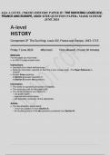
,Neuronal migration
• Chemoattractants and chemorepellants decide whether will or will not grow towards a cell
• Migration can be radial, tangential, or free
• Radial: central neurons move along the long unbranched processes of radial glial cells
o Each glial cell has one basal endfoot in the ventricular zone at the apical surface and
processes that terminate in multiple end-feet at the pial surface
o A neurons leading process wraps around the shaft of the radial glial cell and its
nucleus translocates within the cytoplasm of the leading process
o Integrins promote neural extension
• Tangential: central neurons use axonal tracts as their guides
o The axons of cortical projection neurons reach the internal capsules just as migratory
neurons begin to enter the neocortex; at this intersection immigrating neurons are
tightly associated with the bundles of axons that leave the cortex
• Free: occurs in the PNS without radial glia or axonal tracts
• Growing tip of a neurite is called a growth cone → specialized to identify an appropriate path
for neurite elongation
• Leading edge of the growth cone consists of lamellipodia and extending from these are
filopodia
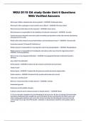
, • Neuroblasts are formed by the asymmetric division of radial
glial cells; neurogenesis can only take place when neural stem
cells have transitioned into radial glial cells
• Proliferating progenitor cells give rise to more differentiated
progeny, but themselves remain in the cell cycle and are
called neural stem cells → in adults in the hippocampus
• Notch signalling regulates the fate of cells in the developing
cerebral cortex
o Neuronal differentiation occurs first, followed by
astrocyte differentiation that peaks at about the time
of birth
o Oligodendrocytes are the last cells to differentiate
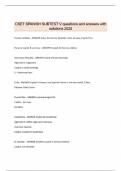
Synapse formation
1. Dendritic filopodium contacts an axon
2. Contact leads to the recruitment of synaptic vesicles and
active zone proteins to the presynaptic membrane
3. Neurotransmitter receptors accumulate post-synaptically
Neuromuscular junction
• Composed of motor neuron, myofiber and Schwann cell; signal by releasing acetylcholine
CNS synapse formation
• Regulated by glutamate and NMDA receptors
• NMDA receptor initiates synaptogenesis through activation of downstream products
Analogies CNS and neuromuscular synapse formation
• Overall structure similarities
• Bi-directional signalling
• Clustering of neurotransmitter receptors
• Synaptic vesicles have similar components
• Synapse elimination during development
Differences CNS and neuromuscular synapse formation
• Central synapses have no basal lamina and no junctional folds, but dendritic spines
• Multiple innervation is common in central synapses
• Difference in neurotransmitters (CNS: excitatory → glutamate, inhibitory → GABA and
glycine)
• Different neurotransmitter receptors
Survival of neurons – neurotrophic factor hypothesis
• Neurons extend their axons to target cells, which secrete low levels of neurotrophic factors
• Neurotrophic factor binds to specific receptors and is internalized and transported to the cell
body, where it promotes neuronal survival
• Neurons that fail to receive adequate amounts of neurotrophic factor die through a program
of cell death termed apoptosis
• Nerve growth factor is a trophic factor promoting neuronal survival is also called the
neurotrophins
• Neurotrophins act as cell surface receptors called trk receptors
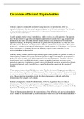
Synaptic plasticity
• Long-term depression
o Brief activation of an excitatory pathway can produce LTD → induced by a minimum
level of postsynaptic depolarization and simultaneous increase in the intracellular
calcium concentration at the postsynaptic neuron
• Long-term potentiation
, o LTP is an increase in synaptic response following potentiating pulses of electrical
stimuli that sustains at a level above the baseline response for hours or longer
o Repeatedly activating the synapse makes it stronger
• Synaptic plasticity can change the amount of neurotransmitter released or the number of
postsynaptic receptors
• NMDA, GABA, AMPA
• Neurons that fire together wire together: presynaptic axon is active at the same time, the
postsynaptic neuron is strongly activated under the influence of other inputs, then the
synapse formed by the presynaptic axon is strengthened
• Neurons that fire out of sync lose their link: when the presynaptic axon is activated and, at
the same time, the postsynaptic neuron is weakly activated by other inputs, then the synapse
formed by the presynaptic axon is weakened
Damage to the NS
Peripheral
• Distal segment experiences Wallerian degeneration
• Endoneurium remains and is preserved by the band of Bünger → column of Schwann cells
Central
• Glial scar forms and glia produce factors that inhibit remyelination and axon repair
• Slower degeneration of the distal segment → axons cannot grow across glial scar
Response in nearby cells
• PNS: Schwann cells break the myelin into small fragments and engulf it and secrete factors
that recruit macrophages that assist in the disposal of debris. Schwann cells also produce
growth factors that promote regeneration of axons
• CNS: myelin forming oligodendrocytes have little ability to dispose myelin and BBB prevents
entry of macrophages, thus clearance depends on microglia → more slowly
Regeneration
Case 01: The developing Nervous System + Lecture Postnatal Brain Development
Prenatal brain development
• Ectoderm will form the central and peripheral nervous system
• Neural plate folds into the neural tube through neurulation and the caudal region will give
rise to the spinal cord and the cranial region will become the brain
• Peripheral nervous system arises from the neural crest
• Three major stages
1. Cell proliferation
2. Cell migration
3. Cell differentiation
Determination and regulation fate of neural cells
Neural induction is mediated by peptide growth factors and their inhibitors
• “die” fate is prevented by signals from neighbouring ectodermal cells → so actually de
inducer is a de-repressor: you always have a die fate, unless the die signal is repressed
• Ectodermal cells can synthesize and secrete BMP, which acts through serine/threonine
kinase class receptors on ectodermal cells and suppress the potential for neural
differentiation and promote epidermal differentiation
• Neural overproduction: approximately half of the neurons generated during embryonic
development will die: cells near the target of a neuron secrete an essential nutrient or
trophic factor which is needed for the survival of the neuron
• Neurotrophins interact with Trk (survival) and p75 (death) receptors
• Main neurotrophins: NGF, brain derived neurotrophic factor (BDNF), and neurotrophin-3
(NT-3)
• Trk: neuronal differentiation largely by the mitogen activated kinase (MAPK) enzymatic
pathways and survival by the phosphatidylinositol-3 kinase pathway
• P75: pathway involves activation of the NF-kappaB, in absence of neurotrophins, p75
receptors are activated
Generation/migration/differentiation of neural and glial cells
• Early neural progenitor cells are capable of self-renewal and give rise to differentiated neural
and glial cells
• Two modes of cell division of the stem cells
o Asymmetric (horizontal): one differentiated daughter cell and one daughter that
keeps stem cell properties
o Symmetric (vertical): two stem cells → expand population of proliferative progenitor
cells
Radial glial cells serve as neural progenitors and structural scaffolds
• Radial glial cells are progenitor cells and can generate everything
• Cell bodies of radial glial cells are located in the ventricular zone and their long processes
extend to the pial surface
• When the generation of neurons is complete, they differentiate into astrocytes: serve as a
scaffold for the migration of neurons that emerge from the ventricular zone
• They generate neurons and astrocytes besides the role in migration and might also serve as
progenitors of neurons in the adult CNS
,Neuronal migration
• Chemoattractants and chemorepellants decide whether will or will not grow towards a cell
• Migration can be radial, tangential, or free
• Radial: central neurons move along the long unbranched processes of radial glial cells
o Each glial cell has one basal endfoot in the ventricular zone at the apical surface and
processes that terminate in multiple end-feet at the pial surface
o A neurons leading process wraps around the shaft of the radial glial cell and its
nucleus translocates within the cytoplasm of the leading process
o Integrins promote neural extension
• Tangential: central neurons use axonal tracts as their guides
o The axons of cortical projection neurons reach the internal capsules just as migratory
neurons begin to enter the neocortex; at this intersection immigrating neurons are
tightly associated with the bundles of axons that leave the cortex
• Free: occurs in the PNS without radial glia or axonal tracts
• Growing tip of a neurite is called a growth cone → specialized to identify an appropriate path
for neurite elongation
• Leading edge of the growth cone consists of lamellipodia and extending from these are
filopodia
, • Neuroblasts are formed by the asymmetric division of radial
glial cells; neurogenesis can only take place when neural stem
cells have transitioned into radial glial cells
• Proliferating progenitor cells give rise to more differentiated
progeny, but themselves remain in the cell cycle and are
called neural stem cells → in adults in the hippocampus
• Notch signalling regulates the fate of cells in the developing
cerebral cortex
o Neuronal differentiation occurs first, followed by
astrocyte differentiation that peaks at about the time
of birth
o Oligodendrocytes are the last cells to differentiate
Synapse formation
1. Dendritic filopodium contacts an axon
2. Contact leads to the recruitment of synaptic vesicles and
active zone proteins to the presynaptic membrane
3. Neurotransmitter receptors accumulate post-synaptically
Neuromuscular junction
• Composed of motor neuron, myofiber and Schwann cell; signal by releasing acetylcholine
CNS synapse formation
• Regulated by glutamate and NMDA receptors
• NMDA receptor initiates synaptogenesis through activation of downstream products
Analogies CNS and neuromuscular synapse formation
• Overall structure similarities
• Bi-directional signalling
• Clustering of neurotransmitter receptors
• Synaptic vesicles have similar components
• Synapse elimination during development
Differences CNS and neuromuscular synapse formation
• Central synapses have no basal lamina and no junctional folds, but dendritic spines
• Multiple innervation is common in central synapses
• Difference in neurotransmitters (CNS: excitatory → glutamate, inhibitory → GABA and
glycine)
• Different neurotransmitter receptors
Survival of neurons – neurotrophic factor hypothesis
• Neurons extend their axons to target cells, which secrete low levels of neurotrophic factors
• Neurotrophic factor binds to specific receptors and is internalized and transported to the cell
body, where it promotes neuronal survival
• Neurons that fail to receive adequate amounts of neurotrophic factor die through a program
of cell death termed apoptosis
• Nerve growth factor is a trophic factor promoting neuronal survival is also called the
neurotrophins
• Neurotrophins act as cell surface receptors called trk receptors
Synaptic plasticity
• Long-term depression
o Brief activation of an excitatory pathway can produce LTD → induced by a minimum
level of postsynaptic depolarization and simultaneous increase in the intracellular
calcium concentration at the postsynaptic neuron
• Long-term potentiation
, o LTP is an increase in synaptic response following potentiating pulses of electrical
stimuli that sustains at a level above the baseline response for hours or longer
o Repeatedly activating the synapse makes it stronger
• Synaptic plasticity can change the amount of neurotransmitter released or the number of
postsynaptic receptors
• NMDA, GABA, AMPA
• Neurons that fire together wire together: presynaptic axon is active at the same time, the
postsynaptic neuron is strongly activated under the influence of other inputs, then the
synapse formed by the presynaptic axon is strengthened
• Neurons that fire out of sync lose their link: when the presynaptic axon is activated and, at
the same time, the postsynaptic neuron is weakly activated by other inputs, then the synapse
formed by the presynaptic axon is weakened
Damage to the NS
Peripheral
• Distal segment experiences Wallerian degeneration
• Endoneurium remains and is preserved by the band of Bünger → column of Schwann cells
Central
• Glial scar forms and glia produce factors that inhibit remyelination and axon repair
• Slower degeneration of the distal segment → axons cannot grow across glial scar
Response in nearby cells
• PNS: Schwann cells break the myelin into small fragments and engulf it and secrete factors
that recruit macrophages that assist in the disposal of debris. Schwann cells also produce
growth factors that promote regeneration of axons
• CNS: myelin forming oligodendrocytes have little ability to dispose myelin and BBB prevents
entry of macrophages, thus clearance depends on microglia → more slowly
Regeneration