Thoraxpathologie: thema ademhaling
Inhoudsopgave
1. INLEIDING ............................................................................................................................ 2
1.1 MACROSCOPIE ................................................................................................................. 2
1.2 MICROSCOPIE .................................................................................................................. 2
1.3 BRONCHO-ALVEOLAIRE BARRIERE .......................................................................................... 2
1.4 HET AFWEERSYSTEEM......................................................................................................... 3
2. INFECTIES ............................................................................................................................. 3
2.1 ALGEMEEN ...................................................................................................................... 3
3. NEOPLASIEËN VAN LONG EN MEDIASTINUM ......................................................................... 5
3.1 CLASSIFICATIE (WHO 2021) ............................................................................................... 5
3.2 ADENOCARCINOOM .......................................................................................................... 6
3.3 SPINOCELLULAIR CARCINOOM .............................................................................................. 6
3.4 NEURO ENDOCRIENE TUMOREN: KLEINCELLIG CARCINOOM ......................................................... 6
3.5 NEURO ENDOCRIENE TUMOREN: GROOTCELLIG CARCINOOM ....................................................... 6
3.6 TYPISCH CARCINOID .......................................................................................................... 7
3.7 ATYPISCH CARCINOID......................................................................................................... 7
3.8 GROOTCELLIG CARCINOOM ................................................................................................. 7
3.9 METASTASEN ................................................................................................................... 7
3.10 IMMUNOHISTOCHEMIE ...................................................................................................... 7
3.11 MOLECULAIRE BIOLOGIE ..................................................................................................... 8
3.12 HAMARTOCHONDROOM..................................................................................................... 9
4. PLEURALE AANDOENINGEN ...................................................................................................... 9
4.1 PLEURA: ANATOMIE ..................................................................................................................... 9
4.2 PLEURALE EFFUSIE........................................................................................................................ 9
4.3 NIET TUMORALE PATHOLOGIE ....................................................................................................... 10
4.4 MALIGNE TUMOR: MESOTHELIOOM................................................................................................ 11
4.5 PLEURALE METASTASEN ............................................................................................................... 11
4.6 DIFFERENTIAAL DIAGNOSE ........................................................................................................... 11
4.7 MALIGNE PLEURAVOCHT .............................................................................................................. 11
4.8 GELOCALISEERDE FIBREUZE PLEURATUMOR....................................................................................... 12
5. OBSTRUCTIEVE LONGZIEKTEN ............................................................................................. 12
5.1 ASTHMA: ...................................................................................................................... 12
5.2 COPD CHRONIC OBSTRUCTIVE PULMONARY DISEASE............................................................... 13
,5.3 BRONCHIËCTASIEËN ......................................................................................................... 13
6. INTERSTITIEEL LONGLIJDEN ................................................................................................. 14
6.1 IDIOPATHISCH INTERSTITIEEL LONGLIJDEN .............................................................................. 14
6.2 ANDERE: ...................................................................................................................... 15
6.3 INTERSTITIEEL LONGLIJDEN: EINDSTADIUM............................................................................. 16
7. PULMONALE CIRCULATIE .................................................................................................... 16
7.1 LONGEMBOLIE ............................................................................................................... 16
VRAGEN ..................................................................................................................................... 16
1. Inleiding
1.1 Macroscopie
- Gladde oppervlakte bekleed met viscerale pleura, rozig longweefsel, zwarte puntjes
komen door stof, iedereen heeft dit
- 3 loben aan de rechter kant, normaal gewicht: 400-450 g 2
- loben aan de linker kant, normaal gewicht: 300-350 g
- Lobben/kwabben > segmenten > lobulus > acinus > alveolen
o Lobuli: piramidaal, basis tegen pleura en apex naar longhillus
o Acini: piramidaal, basis naar rande lobule, apex naar bronchiolen, ( bestaat uit
resp bronchiolen,alv sacculi, alveolen
o Pathologie: centrolobulair en/of subpleuraal/subseptaal
- Trachea> hoofdBronchi > secundaire bronchi> bronchiolen (geen kraakbeen) 1e,2e, 3e
orde( terminale bronchiolen) > alveolaire ductus
- Dubbele arteriële bloedsomloop: van a pulm (zuurstofarm, langs bronchiolen) +
bronchiale arteries ( zuurstofrijk, aftakking aorta, langs bronchiolen)
Veneuze afvloei : zuurstofrijk, naar pulm venen, langs septa
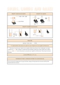
1.2 Microscopie
- Bronchus: trilhaarcellen & slijmbekercellen & neuroendocriene cellen & basale cellen:
pseudogestratifieerd gecillieerd resp epitheel; basale laag; stroma; bronchiale klieren;
glad spierweefsel (bij bronchiolen meer aanw, kunnen sterk vernauwen bij
bronchoconstrictie); kraakbeen ( bij trachea en bronchi)
- Perifeer longparenchym: alveoli, holtes met elkaar verbonden, interalveolaire septa
(pneumocyten) tussen alveolen zitten ook cappilairen
1.3 broncho-alveolaire barriere
- binnenkant alveoli: surfactant, lippoproteine materiaal, aanmaak door type II
pneumocyten
- epitheliale opp: pneumocyten bekleden interalveolaire septa
o pneumocyten type I: zeer gevoelig aan aggressies, knn necroseren
o pneumocyten type II: regeneratie van type I pneumocyten
, - basale membraan ad epitheel
- stroma: enkele elastische collageenvezels en histocyten
- basale membraan ad endotheel
- cappilair endotheel met RBC in cappilair
1.4 het afweersysteem
- waarom:
o Erg grote epitheliale oppervlakte: direct contact met de buitenlucht
o Inademen naast zuurstof van fijnstof, toxische gassen en micro- organismen
o Inoculatie van bacteriën vanuit de naso-/oro-farynx
- Al deze elementen moeten verwijderd worden zonder een inadequate inflammatoire
reactie uit te lokken
- In de bovenste luchtwegen en bronchi:
o Mucociliaire clearance van de neus en de tracheo-bronchiale boom
geassocieerd met hoest dmv trilhaarcellen die mucus evacueren
o Slijmvlies van de luchtwegen, die een fysieke barrière zijn en ook defensie
moleculen produceren
o Lymfocyten van het lymfoïd weefsel geassocieerd aan de bronchi (BALT)
o Productie van een hoge concentratie immuunglobuline A, die toxines, virussen
en bacteriën neutraliseert
- In de alveoli
o Histiocyten
o Immunoglobulines van het broncho-alveolair vocht (surfactant)
o Lymfocyten (10%)
o Neutrofielen (2%) en eosinofielen
o → altijd een paar inflammatoire cellen aanwezig in alveolair vocht dit wil niet
zeggen dat er altijd inflammatie aanw is! → verhouding versch elementen
2. Infecties
2.1 algemeen
- Definitie: Inflammatie die het hele longweefsel kan aantasten: longparenchym en
bronchi
- Classificatie
o Volgens de anatomie
▪ Lobaire pneumonie
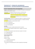
▪ Bronchopneumonie: infectie rond
brochovasculaire bundels, centraal in
bronchus en omringende alv structuren
(macroscopisch: kleine puntjes zichtbaar
met ertussen normaal longweefsel)
o Volgens origine:
▪ Primair: door inhalatie of aspiratie van oro-faryngeale
gecontamineerde secreties
▪ Secundair: door hematogene disseminatie afkomstig van een ander
orgaan
- Bacteriële longinfectie: lobaire pneumonie
o Aantasting van een gehele longkwab
Inhoudsopgave
1. INLEIDING ............................................................................................................................ 2
1.1 MACROSCOPIE ................................................................................................................. 2
1.2 MICROSCOPIE .................................................................................................................. 2
1.3 BRONCHO-ALVEOLAIRE BARRIERE .......................................................................................... 2
1.4 HET AFWEERSYSTEEM......................................................................................................... 3
2. INFECTIES ............................................................................................................................. 3
2.1 ALGEMEEN ...................................................................................................................... 3
3. NEOPLASIEËN VAN LONG EN MEDIASTINUM ......................................................................... 5
3.1 CLASSIFICATIE (WHO 2021) ............................................................................................... 5
3.2 ADENOCARCINOOM .......................................................................................................... 6
3.3 SPINOCELLULAIR CARCINOOM .............................................................................................. 6
3.4 NEURO ENDOCRIENE TUMOREN: KLEINCELLIG CARCINOOM ......................................................... 6
3.5 NEURO ENDOCRIENE TUMOREN: GROOTCELLIG CARCINOOM ....................................................... 6
3.6 TYPISCH CARCINOID .......................................................................................................... 7
3.7 ATYPISCH CARCINOID......................................................................................................... 7
3.8 GROOTCELLIG CARCINOOM ................................................................................................. 7
3.9 METASTASEN ................................................................................................................... 7
3.10 IMMUNOHISTOCHEMIE ...................................................................................................... 7
3.11 MOLECULAIRE BIOLOGIE ..................................................................................................... 8
3.12 HAMARTOCHONDROOM..................................................................................................... 9
4. PLEURALE AANDOENINGEN ...................................................................................................... 9
4.1 PLEURA: ANATOMIE ..................................................................................................................... 9
4.2 PLEURALE EFFUSIE........................................................................................................................ 9
4.3 NIET TUMORALE PATHOLOGIE ....................................................................................................... 10
4.4 MALIGNE TUMOR: MESOTHELIOOM................................................................................................ 11
4.5 PLEURALE METASTASEN ............................................................................................................... 11
4.6 DIFFERENTIAAL DIAGNOSE ........................................................................................................... 11
4.7 MALIGNE PLEURAVOCHT .............................................................................................................. 11
4.8 GELOCALISEERDE FIBREUZE PLEURATUMOR....................................................................................... 12
5. OBSTRUCTIEVE LONGZIEKTEN ............................................................................................. 12
5.1 ASTHMA: ...................................................................................................................... 12
5.2 COPD CHRONIC OBSTRUCTIVE PULMONARY DISEASE............................................................... 13
,5.3 BRONCHIËCTASIEËN ......................................................................................................... 13
6. INTERSTITIEEL LONGLIJDEN ................................................................................................. 14
6.1 IDIOPATHISCH INTERSTITIEEL LONGLIJDEN .............................................................................. 14
6.2 ANDERE: ...................................................................................................................... 15
6.3 INTERSTITIEEL LONGLIJDEN: EINDSTADIUM............................................................................. 16
7. PULMONALE CIRCULATIE .................................................................................................... 16
7.1 LONGEMBOLIE ............................................................................................................... 16
VRAGEN ..................................................................................................................................... 16
1. Inleiding
1.1 Macroscopie
- Gladde oppervlakte bekleed met viscerale pleura, rozig longweefsel, zwarte puntjes
komen door stof, iedereen heeft dit
- 3 loben aan de rechter kant, normaal gewicht: 400-450 g 2
- loben aan de linker kant, normaal gewicht: 300-350 g
- Lobben/kwabben > segmenten > lobulus > acinus > alveolen
o Lobuli: piramidaal, basis tegen pleura en apex naar longhillus
o Acini: piramidaal, basis naar rande lobule, apex naar bronchiolen, ( bestaat uit
resp bronchiolen,alv sacculi, alveolen
o Pathologie: centrolobulair en/of subpleuraal/subseptaal
- Trachea> hoofdBronchi > secundaire bronchi> bronchiolen (geen kraakbeen) 1e,2e, 3e
orde( terminale bronchiolen) > alveolaire ductus
- Dubbele arteriële bloedsomloop: van a pulm (zuurstofarm, langs bronchiolen) +
bronchiale arteries ( zuurstofrijk, aftakking aorta, langs bronchiolen)
Veneuze afvloei : zuurstofrijk, naar pulm venen, langs septa
1.2 Microscopie
- Bronchus: trilhaarcellen & slijmbekercellen & neuroendocriene cellen & basale cellen:
pseudogestratifieerd gecillieerd resp epitheel; basale laag; stroma; bronchiale klieren;
glad spierweefsel (bij bronchiolen meer aanw, kunnen sterk vernauwen bij
bronchoconstrictie); kraakbeen ( bij trachea en bronchi)
- Perifeer longparenchym: alveoli, holtes met elkaar verbonden, interalveolaire septa
(pneumocyten) tussen alveolen zitten ook cappilairen
1.3 broncho-alveolaire barriere
- binnenkant alveoli: surfactant, lippoproteine materiaal, aanmaak door type II
pneumocyten
- epitheliale opp: pneumocyten bekleden interalveolaire septa
o pneumocyten type I: zeer gevoelig aan aggressies, knn necroseren
o pneumocyten type II: regeneratie van type I pneumocyten
, - basale membraan ad epitheel
- stroma: enkele elastische collageenvezels en histocyten
- basale membraan ad endotheel
- cappilair endotheel met RBC in cappilair
1.4 het afweersysteem
- waarom:
o Erg grote epitheliale oppervlakte: direct contact met de buitenlucht
o Inademen naast zuurstof van fijnstof, toxische gassen en micro- organismen
o Inoculatie van bacteriën vanuit de naso-/oro-farynx
- Al deze elementen moeten verwijderd worden zonder een inadequate inflammatoire
reactie uit te lokken
- In de bovenste luchtwegen en bronchi:
o Mucociliaire clearance van de neus en de tracheo-bronchiale boom
geassocieerd met hoest dmv trilhaarcellen die mucus evacueren
o Slijmvlies van de luchtwegen, die een fysieke barrière zijn en ook defensie
moleculen produceren
o Lymfocyten van het lymfoïd weefsel geassocieerd aan de bronchi (BALT)
o Productie van een hoge concentratie immuunglobuline A, die toxines, virussen
en bacteriën neutraliseert
- In de alveoli
o Histiocyten
o Immunoglobulines van het broncho-alveolair vocht (surfactant)
o Lymfocyten (10%)
o Neutrofielen (2%) en eosinofielen
o → altijd een paar inflammatoire cellen aanwezig in alveolair vocht dit wil niet
zeggen dat er altijd inflammatie aanw is! → verhouding versch elementen
2. Infecties
2.1 algemeen
- Definitie: Inflammatie die het hele longweefsel kan aantasten: longparenchym en
bronchi
- Classificatie
o Volgens de anatomie
▪ Lobaire pneumonie
▪ Bronchopneumonie: infectie rond
brochovasculaire bundels, centraal in
bronchus en omringende alv structuren
(macroscopisch: kleine puntjes zichtbaar
met ertussen normaal longweefsel)
o Volgens origine:
▪ Primair: door inhalatie of aspiratie van oro-faryngeale
gecontamineerde secreties
▪ Secundair: door hematogene disseminatie afkomstig van een ander
orgaan
- Bacteriële longinfectie: lobaire pneumonie
o Aantasting van een gehele longkwab