The Digestive System
Friday, 21 November 2025 16:59
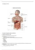
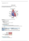
Digestive system anatomy
The organs of the digestive system are divided into:
1. Alimentary Canal (GI Tract / Gut)
• A continuous muscular tube that runs from mouth to anus.
• Main functions:
→ Digestion: Breaks food into smaller molecules.
→ Absorption: Moves these molecules through the epithelial lining into the blood.
• Organs included:
→ Mouth
→ Pharynx
→ Esophagus
→ Stomach
→ Small intestine
→ Large intestine
• Food in this tube is technically outside the body until absorbed, because the GI tract opens to the external environment
at both ends.
• It is approximately 9 meters in a cadaver (muscles relaxed).
→ Shorter in living people due to muscle tone.
• Layers of the alimentary canal:
1. Mucosa
• Innermost layer (lines the lumen).
• Functions include secretion, absorption and protection.
, at both ends.
• It is approximately 9 meters in a cadaver (muscles relaxed).
→ Shorter in living people due to muscle tone.
• Layers of the alimentary canal:
1. Mucosa
• Innermost layer (lines the lumen).
• Functions include secretion, absorption and protection.
• Has three sublayers:
► Epithelium
• Simple columnar epithelium for most of the GI tract
• Stratified squamous in mouth, esophagus, pharynx and anus for protection from abrasion.
• Contains mucus-secreting cells, enzyme-producing cells and hormone-producing enteroendocrine cells
→ GI mucosa also acts as a diffuse endocrine organ.
► Lamina propria
• Loose areolar connective tissue.
• Contain capillaries which nourish epithelium & absorb nutrients
• Contains lymphoid follicles for immune defense.
→ Part of MALT (mucosa-associated lymphoid tissue).
→ Large collections of lymphoid follicles occur within the pharynx (as the tonsils) and in the appendix.
► Muscularis mucosae
• Thin layer of smooth muscle
• Creates local movements of mucosa
• In small intestine it forms folds to increase surface area.
2. Submucosa
• Second layer.
• Consists of areolar connective tissue.
• Contains:
→ Blood vessels
→ Lymphatic vessels
→ Lymphoid follicles
→ Nerve fibers (submucosal plexus / Meissner’s plexus)
→ Elastic fibers: allow stomach to stretch and recoil after meals.
3. Muscularis Externa (Muscularis)
• Smooth muscle layer responsible for motility.
• Contains two layers:
→ Inner circular layer
• Circular layer can thicken to form sphincters which regulate passage and prevent backflow.
→ Outer longitudinal layer
→ The stomach contains a third layer, called the oblique layer (inner layer).
→ Enhances mixing, churning, and propulsion of food.
• Functions:
• Segmentation (mixing)
• Peristalsis (propulsion)
• Contains myenteric plexus (Auerbach’s), which controls motility.
4. Serosa (visceral peritoneum)
► Outermost layer for intraperitoneal organs.
► Consists of areolar connective tissue covered by mesothelium (simple squamous epithelial cells)
→ Exceptions:
► In the esophagus, serosa is replaced by adventitia (fibrous connective tissue)
→ It anchors esophagus in thorax.
→ Retroperitoneal organs have both serosa and adventitia.
2. Accessory Digestive Organs
• These assist with digestion but are not part of the tube itself.
• Function:
→ They make secretions (saliva, bile, digestive enzymes) that are delivered to the GI tract through ducts to help break
down food.
• Accessory structures include:
→ Teeth & Tongue
→ Gallbladder
→ Digestive glands:
→ Salivary glands
→ Liver
→ Pancreas
, down food.
• Accessory structures include:
→ Teeth & Tongue
→ Gallbladder
→ Digestive glands:
→ Salivary glands
→ Liver
→ Pancreas
Intraperitoneal vs. Retroperitoneal Organs
► Retroperitoneal organs
• During development, some organs lose their mesentery and become stuck to the posterior body wall.
• Located behind the peritoneum, so only have anterior peritoneal covering.
• Examples include:
• Most of the pancreas
• Most of the duodenum
• Parts of the large intestine (ascending, descending colon)
► Intraperitoneal organs
These keep their mesentery and remain suspended in the peritoneal cavity.
• Examples include:
→ Stomach
→ Jejunum
→ Ileum
→ Transverse colon
→ Liver
• The peritoneum: serous membrane that lines the abdominal cavity and covers most abdominal organs.
→ Most digestive organs sit inside the abdominopelvic cavity.
→ It has two layers:
1. Visceral peritoneum: covers the external surfaces of most digestive organs.
2. Parietal peritoneum: lines the body wall.
→ Between them is the peritoneal cavity.
→ Contains serous fluid, which reduces friction in organs during digestion.
→ Mesentery: a double layer of peritoneum, that extends to the digestive organs from the body wall.
→ Functions:
• Provides pathways for blood vessels, lymphatics and nerves.
• Anchors organs in place.
• Stores fat.
→ In most places the mesentery is dorsal (attaches to posterior abdominal wall), but there are ventral mesenteries too
(such as the one that extends from the liver to the anterior abdominal wall).
---------------------------------------------------------------------------------------------------
Processes within digestion
1. Ingestion
• Taking food into the digestive tract, through the mouth.
2. Propulsion
• Moving food through the GI tract.
• Includes:
→ Swallowing (deglutition)(in
→ Peristalsis – involuntary.
• Alternating waves of contraction and relaxation, inorder to push food forward.
• Occurs in the muscular walls of GI organs.
• Strong enough to move food even if you’re upside down.
• Occurs in esophagus, stomach, small and large intestine.
• Process of swallowing:
1) Buccal Phase (Voluntary)
• Occurs in the mouth
• Tongue contracts forcing bolus into the oropharynx
• Once the bolus touches the back of the pharynx a reflex is triggered
→ Control passes from voluntary to involuntary.
2) Pharyngeal – Esophageal Phase (Involuntary)
• Controlled by the swallowing center in the medulla and lower pons.
• Signals travel via cranial nerves (especially the vagus nerve).
• Breathing stops momentarily, to prevent aspiration.
, • Occurs in the mouth
• Tongue contracts forcing bolus into the oropharynx
• Once the bolus touches the back of the pharynx a reflex is triggered
→ Control passes from voluntary to involuntary.
2) Pharyngeal – Esophageal Phase (Involuntary)
• Controlled by the swallowing center in the medulla and lower pons.
• Signals travel via cranial nerves (especially the vagus nerve).
• Breathing stops momentarily, to prevent aspiration.
3. Mechanical Breakdown
• Physically breaking food into smaller pieces to increase surface area for enzymes.
• Includes:
→ Chewing (mastication).
→ Mixing food with saliva by the tongue.
→ Churning in the stomach.
→ Segmentation in the small intestine.
→ Rhythmic contractions that mix food with digestive juices
→ Helps expose food repeatedly to the absorptive surface, leading to better absorption.
→ Sometimes called “mechanical digestion,” but technically digestion is chemical breakdown by enzymes.
4. Digestion
• Chemical breakdown of complex food molecules into basic building blocks using enzymes.
→ Occurs in the lumen of the GI tract.
• Examples:
→ Proteins → amino acids
→ Carbohydrates → monosaccharides
→ Fats → fatty acids + glycerol
5. Absorption
• Moving digested nutrients through mucosal cells into blood or lymph.
• Substances that get absorbed include:
→ Monomers (amino acids, sugars, fatty acids)
→ Vitamins
→ Minerals
→ Water
• Occurs mostly in small intestine, with some water and ions being absorbed in large intestine.
6. Defecation
• Eliminating indigestible material (feces) from the GI tract through the anus.
• Involves the large intestine and anus.
→ Defecation reflex:
1. Rectal filling:
→ Feces enter the rectum from the sigmoid colon.
→ Stretch receptors in the rectal wall detect distension.
2. Signals are sent to the sacral spinal cord via parasympathetic nerves.
3. Reflex activation:
→ Contraction of the rectum and sigmoid colon (to push feces downward)
→ Relaxation of the internal anal sphincter (involuntary smooth muscle)
4. Voluntary control:
→ Signals reach the brain, allowing conscious control of the external anal sphincter (skeletal muscle).
5. Completion:
→ When ready, voluntary contraction of abdominal muscles and diaphragm (Valsalva maneuver) increases intra-
abdominal pressure to help expel feces.
→ The levator ani lifts the anal canal to assist in smooth evacuation.
Friday, 21 November 2025 16:59
Digestive system anatomy
The organs of the digestive system are divided into:
1. Alimentary Canal (GI Tract / Gut)
• A continuous muscular tube that runs from mouth to anus.
• Main functions:
→ Digestion: Breaks food into smaller molecules.
→ Absorption: Moves these molecules through the epithelial lining into the blood.
• Organs included:
→ Mouth
→ Pharynx
→ Esophagus
→ Stomach
→ Small intestine
→ Large intestine
• Food in this tube is technically outside the body until absorbed, because the GI tract opens to the external environment
at both ends.
• It is approximately 9 meters in a cadaver (muscles relaxed).
→ Shorter in living people due to muscle tone.
• Layers of the alimentary canal:
1. Mucosa
• Innermost layer (lines the lumen).
• Functions include secretion, absorption and protection.
, at both ends.
• It is approximately 9 meters in a cadaver (muscles relaxed).
→ Shorter in living people due to muscle tone.
• Layers of the alimentary canal:
1. Mucosa
• Innermost layer (lines the lumen).
• Functions include secretion, absorption and protection.
• Has three sublayers:
► Epithelium
• Simple columnar epithelium for most of the GI tract
• Stratified squamous in mouth, esophagus, pharynx and anus for protection from abrasion.
• Contains mucus-secreting cells, enzyme-producing cells and hormone-producing enteroendocrine cells
→ GI mucosa also acts as a diffuse endocrine organ.
► Lamina propria
• Loose areolar connective tissue.
• Contain capillaries which nourish epithelium & absorb nutrients
• Contains lymphoid follicles for immune defense.
→ Part of MALT (mucosa-associated lymphoid tissue).
→ Large collections of lymphoid follicles occur within the pharynx (as the tonsils) and in the appendix.
► Muscularis mucosae
• Thin layer of smooth muscle
• Creates local movements of mucosa
• In small intestine it forms folds to increase surface area.
2. Submucosa
• Second layer.
• Consists of areolar connective tissue.
• Contains:
→ Blood vessels
→ Lymphatic vessels
→ Lymphoid follicles
→ Nerve fibers (submucosal plexus / Meissner’s plexus)
→ Elastic fibers: allow stomach to stretch and recoil after meals.
3. Muscularis Externa (Muscularis)
• Smooth muscle layer responsible for motility.
• Contains two layers:
→ Inner circular layer
• Circular layer can thicken to form sphincters which regulate passage and prevent backflow.
→ Outer longitudinal layer
→ The stomach contains a third layer, called the oblique layer (inner layer).
→ Enhances mixing, churning, and propulsion of food.
• Functions:
• Segmentation (mixing)
• Peristalsis (propulsion)
• Contains myenteric plexus (Auerbach’s), which controls motility.
4. Serosa (visceral peritoneum)
► Outermost layer for intraperitoneal organs.
► Consists of areolar connective tissue covered by mesothelium (simple squamous epithelial cells)
→ Exceptions:
► In the esophagus, serosa is replaced by adventitia (fibrous connective tissue)
→ It anchors esophagus in thorax.
→ Retroperitoneal organs have both serosa and adventitia.
2. Accessory Digestive Organs
• These assist with digestion but are not part of the tube itself.
• Function:
→ They make secretions (saliva, bile, digestive enzymes) that are delivered to the GI tract through ducts to help break
down food.
• Accessory structures include:
→ Teeth & Tongue
→ Gallbladder
→ Digestive glands:
→ Salivary glands
→ Liver
→ Pancreas
, down food.
• Accessory structures include:
→ Teeth & Tongue
→ Gallbladder
→ Digestive glands:
→ Salivary glands
→ Liver
→ Pancreas
Intraperitoneal vs. Retroperitoneal Organs
► Retroperitoneal organs
• During development, some organs lose their mesentery and become stuck to the posterior body wall.
• Located behind the peritoneum, so only have anterior peritoneal covering.
• Examples include:
• Most of the pancreas
• Most of the duodenum
• Parts of the large intestine (ascending, descending colon)
► Intraperitoneal organs
These keep their mesentery and remain suspended in the peritoneal cavity.
• Examples include:
→ Stomach
→ Jejunum
→ Ileum
→ Transverse colon
→ Liver
• The peritoneum: serous membrane that lines the abdominal cavity and covers most abdominal organs.
→ Most digestive organs sit inside the abdominopelvic cavity.
→ It has two layers:
1. Visceral peritoneum: covers the external surfaces of most digestive organs.
2. Parietal peritoneum: lines the body wall.
→ Between them is the peritoneal cavity.
→ Contains serous fluid, which reduces friction in organs during digestion.
→ Mesentery: a double layer of peritoneum, that extends to the digestive organs from the body wall.
→ Functions:
• Provides pathways for blood vessels, lymphatics and nerves.
• Anchors organs in place.
• Stores fat.
→ In most places the mesentery is dorsal (attaches to posterior abdominal wall), but there are ventral mesenteries too
(such as the one that extends from the liver to the anterior abdominal wall).
---------------------------------------------------------------------------------------------------
Processes within digestion
1. Ingestion
• Taking food into the digestive tract, through the mouth.
2. Propulsion
• Moving food through the GI tract.
• Includes:
→ Swallowing (deglutition)(in
→ Peristalsis – involuntary.
• Alternating waves of contraction and relaxation, inorder to push food forward.
• Occurs in the muscular walls of GI organs.
• Strong enough to move food even if you’re upside down.
• Occurs in esophagus, stomach, small and large intestine.
• Process of swallowing:
1) Buccal Phase (Voluntary)
• Occurs in the mouth
• Tongue contracts forcing bolus into the oropharynx
• Once the bolus touches the back of the pharynx a reflex is triggered
→ Control passes from voluntary to involuntary.
2) Pharyngeal – Esophageal Phase (Involuntary)
• Controlled by the swallowing center in the medulla and lower pons.
• Signals travel via cranial nerves (especially the vagus nerve).
• Breathing stops momentarily, to prevent aspiration.
, • Occurs in the mouth
• Tongue contracts forcing bolus into the oropharynx
• Once the bolus touches the back of the pharynx a reflex is triggered
→ Control passes from voluntary to involuntary.
2) Pharyngeal – Esophageal Phase (Involuntary)
• Controlled by the swallowing center in the medulla and lower pons.
• Signals travel via cranial nerves (especially the vagus nerve).
• Breathing stops momentarily, to prevent aspiration.
3. Mechanical Breakdown
• Physically breaking food into smaller pieces to increase surface area for enzymes.
• Includes:
→ Chewing (mastication).
→ Mixing food with saliva by the tongue.
→ Churning in the stomach.
→ Segmentation in the small intestine.
→ Rhythmic contractions that mix food with digestive juices
→ Helps expose food repeatedly to the absorptive surface, leading to better absorption.
→ Sometimes called “mechanical digestion,” but technically digestion is chemical breakdown by enzymes.
4. Digestion
• Chemical breakdown of complex food molecules into basic building blocks using enzymes.
→ Occurs in the lumen of the GI tract.
• Examples:
→ Proteins → amino acids
→ Carbohydrates → monosaccharides
→ Fats → fatty acids + glycerol
5. Absorption
• Moving digested nutrients through mucosal cells into blood or lymph.
• Substances that get absorbed include:
→ Monomers (amino acids, sugars, fatty acids)
→ Vitamins
→ Minerals
→ Water
• Occurs mostly in small intestine, with some water and ions being absorbed in large intestine.
6. Defecation
• Eliminating indigestible material (feces) from the GI tract through the anus.
• Involves the large intestine and anus.
→ Defecation reflex:
1. Rectal filling:
→ Feces enter the rectum from the sigmoid colon.
→ Stretch receptors in the rectal wall detect distension.
2. Signals are sent to the sacral spinal cord via parasympathetic nerves.
3. Reflex activation:
→ Contraction of the rectum and sigmoid colon (to push feces downward)
→ Relaxation of the internal anal sphincter (involuntary smooth muscle)
4. Voluntary control:
→ Signals reach the brain, allowing conscious control of the external anal sphincter (skeletal muscle).
5. Completion:
→ When ready, voluntary contraction of abdominal muscles and diaphragm (Valsalva maneuver) increases intra-
abdominal pressure to help expel feces.
→ The levator ani lifts the anal canal to assist in smooth evacuation.