The Cardiovascular System
Friday, 31 October 2025 09:38
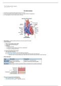
The Heart anatomy
• The heart is a muscular organ about the size of your fist.
• It lies in the thoracic cavity, between the lungs, in a space called the mediastinum
• It is tilted slightly so that the apex points to the left.
• Pericardium: a double-walled sac that surrounds and protects the heart.
→ It has two layers:
1. Fibrous pericardium (outer layer)
→ Tough, dense connective tissue.
→ Functions:
• Protects the heart
• Anchors it to surrounding structures (e.g. diaphragm)
• Prevents overfilling with blood
2. Serous pericardium (inner layer)
→ Thin, two-layer membrane.
► Parietal layer: lines the inside of the fibrous pericardium.
► Visceral layer (epicardium): covers the heart surface and forms part of the heart wall.
→ Between them is the pericardial cavity, filled with serous fluid that reduces friction as the heart beat.
• Heart wall layers:
Layer Description Composition
Endocardium Inner lining of chambers Endothelium + connective tissue
Myocardium Middle, thick muscle layer. Cardiac muscle cells (cardiomyocytes)
Contracts.
Epicardium Visceral layer of serious percardium Serous membrane with connective & fat tissue
• The heart is seperated longitudinally by:
→ The interatrial septum - seperates atria
→ The interventricular septum - seperates ventricles
Cardiac Muscle
• Striated due to sarcomeres
• Contracts via the sliding filament mechanism.
, • The heart is seperated longitudinally by:
→ The interatrial septum - seperates atria
→ The interventricular septum - seperates ventricles
Cardiac Muscle
• Striated due to sarcomeres
• Contracts via the sliding filament mechanism.
• Contains intercalated discs.
→ They contain desmosomes (to prevent sepeartion) and gap junctions (allowing ions to pass).
→ Allow cardiac cells to function as a functional syncytium (act as a single cooridnated unit).
• Contain many mitochondria (25-35% of cell volume), ensuring high fatique resistence.
Chambers of the heart
• Atria
→ Are the receiving chambers.
→ Have thin walls and contract only lightly to push blood into the ventricles, contributing little to overall pumping force.
1. Right Atrium
• Has two parts:
→ Smooth posterior wall
→ Anterior wall with ridged pectinate muscles.
• The two regions are separated by the crista terminalis (a C-shaped ridge).
• The interatrial septum contains the fossa ovalis, a remnant of the fetal foramen ovale.
→ Foramen ovale: opening in the interatrial septum that allows blood to pass directly from the right atrium to the left
atrium, bypassing the fetal lungs (which are not yet functioning before birth).
• Receives deoxygenated blood from three sources:
1. Superior vena cava – from areas above the diaphragm.
2. Inferior vena cava – from areas below the diaphragm.
3. Coronary sinus – from the heart’s myocardium.
2. Left Atrium
• Mostly smooth-walled, with pectinate muscles only in the auricle.
→ Each atrium (right and left) has one auricle.
→ They are wrinkled, mucular protrusions that increase atrial volume.
• Receives oxygenated blood from four pulmonary veins returning from the lungs.
• Forms most of the base of the heart.
• Ventricles
→ Are the pumping chambers.
→ Make up most of its volume.
→ Have thicker walls than atria because they must pump blood with greater force.
→ Internal features:
→ Trabeculae carneae – irregular ridges of muscle on the inner wall.
→ Papillary muscles – cone-shaped projections that help control the heart valves during contraction.
1. Right ventricle
• Pumps deoxygenated blood into the pulmonary trunk (which divides into right and left pulmonery artery), to the lungs for
gas exchange.
2. Left ventricle
• Pumps oxygenated blood into the aorta, to supply all tissues.
Heart Surface Faces Main Chamber
Anterior (sternocostal) Forward Right ventricle
Posterior (base) Backward Left atrium
Inferior (diaphragmatic) Downward Left & right ventricles
, gas exchange.
2. Left ventricle
• Pumps oxygenated blood into the aorta, to supply all tissues.
Heart Surface Faces Main Chamber
Anterior (sternocostal) Forward Right ventricle
Posterior (base) Backward Left atrium
Inferior (diaphragmatic) Downward Left & right ventricles
Valves in the heart
1. Atrioventricular (AV) Valves
• Prevent backflow of blood into the atriums during ventricular contraction.
→ When atria contract: AV valves open, allowing blood to flow into ventricles.
→ When ventricles contract: pressure pushes the valves closed, preventing backflow.
• Papillary muscles contract to hold the valve cusps in place via chordae tendineae, preventing them from inverting.
1. Tricuspid valve
• Between right atrium and right ventricle
• Made up of 3 cusps (flaps).
2. Mitral (bicuspid) valve
• Between left atrium and left ventricle
• Made up of two cusps (flaps).
2. Semilunar (SL) Valves
• Prevents backflow of blood into the right ventricle after contraction.
→ When ventricles contract: pressure forces SL valves open, and blood flows into the pulmonary trunk or aorta.
→ When ventricles relax: pressure in arteries becomes higher, causing SL valves to close.
• Don't contain chordae tendinae.
• Made up of 3 cusps (flaps).
1. Pulmonary valve
→ Between right ventricle and pulmonary trunk.
2. Aortic valve
→ Between left ventricle and aorta.
Blood vessels of the heart
Vessel Type From To Notes
Aorta Artery Left ventricle Body tissues - Largest artery.
- Branches into brachiocephalic, left common carotid, and left
subclavian arteries
- Carries blood under high pressure.
Vena cava Vein Body tissues Right atrium - Two parts: superior (head/arms) and inferior (trunk/legs)
Pulmonary Artery Right ventricle Lungs - Divides into left and right pulmonary arteries.
artery - Only artery that carries deoxygenated blood.
Pulmonary Vein Lungs Left atrium - Four veins (two from each lung).
veins - Only veins that carry oxygenated blood.
--------------------------------------------------------------------------------------------------------------------------------------------------
Pathway of blood through the heart
1. Deoxygenated Blood from the body enters the right atrium, through vena cava.
→ Superior vena cava (SVC) – returns blood from the upper body
→ Inferior vena cava (IVC) – returns blood from the lower body
→ Coronary sinus – returns blood from the heart muscle itself.
2. Blood flows into right ventricle, through tricuspid valve.
3. Blood flows into pulmonary trunk through the pulmonery valve.
4. Pulmonery trunk splits into right and left arteries, which travel to each lung to pick up oxygen.
5. Oxygenated blood enters the left atrium through four pulmonary veins
6. Blood enters the left ventricle, through the mitral valve.
→ Left ventricle contracts.
7. Blood enters the aorta, through the aortic valve.
8. Blood is distributed to the entire body.
Friday, 31 October 2025 09:38
The Heart anatomy
• The heart is a muscular organ about the size of your fist.
• It lies in the thoracic cavity, between the lungs, in a space called the mediastinum
• It is tilted slightly so that the apex points to the left.
• Pericardium: a double-walled sac that surrounds and protects the heart.
→ It has two layers:
1. Fibrous pericardium (outer layer)
→ Tough, dense connective tissue.
→ Functions:
• Protects the heart
• Anchors it to surrounding structures (e.g. diaphragm)
• Prevents overfilling with blood
2. Serous pericardium (inner layer)
→ Thin, two-layer membrane.
► Parietal layer: lines the inside of the fibrous pericardium.
► Visceral layer (epicardium): covers the heart surface and forms part of the heart wall.
→ Between them is the pericardial cavity, filled with serous fluid that reduces friction as the heart beat.
• Heart wall layers:
Layer Description Composition
Endocardium Inner lining of chambers Endothelium + connective tissue
Myocardium Middle, thick muscle layer. Cardiac muscle cells (cardiomyocytes)
Contracts.
Epicardium Visceral layer of serious percardium Serous membrane with connective & fat tissue
• The heart is seperated longitudinally by:
→ The interatrial septum - seperates atria
→ The interventricular septum - seperates ventricles
Cardiac Muscle
• Striated due to sarcomeres
• Contracts via the sliding filament mechanism.
, • The heart is seperated longitudinally by:
→ The interatrial septum - seperates atria
→ The interventricular septum - seperates ventricles
Cardiac Muscle
• Striated due to sarcomeres
• Contracts via the sliding filament mechanism.
• Contains intercalated discs.
→ They contain desmosomes (to prevent sepeartion) and gap junctions (allowing ions to pass).
→ Allow cardiac cells to function as a functional syncytium (act as a single cooridnated unit).
• Contain many mitochondria (25-35% of cell volume), ensuring high fatique resistence.
Chambers of the heart
• Atria
→ Are the receiving chambers.
→ Have thin walls and contract only lightly to push blood into the ventricles, contributing little to overall pumping force.
1. Right Atrium
• Has two parts:
→ Smooth posterior wall
→ Anterior wall with ridged pectinate muscles.
• The two regions are separated by the crista terminalis (a C-shaped ridge).
• The interatrial septum contains the fossa ovalis, a remnant of the fetal foramen ovale.
→ Foramen ovale: opening in the interatrial septum that allows blood to pass directly from the right atrium to the left
atrium, bypassing the fetal lungs (which are not yet functioning before birth).
• Receives deoxygenated blood from three sources:
1. Superior vena cava – from areas above the diaphragm.
2. Inferior vena cava – from areas below the diaphragm.
3. Coronary sinus – from the heart’s myocardium.
2. Left Atrium
• Mostly smooth-walled, with pectinate muscles only in the auricle.
→ Each atrium (right and left) has one auricle.
→ They are wrinkled, mucular protrusions that increase atrial volume.
• Receives oxygenated blood from four pulmonary veins returning from the lungs.
• Forms most of the base of the heart.
• Ventricles
→ Are the pumping chambers.
→ Make up most of its volume.
→ Have thicker walls than atria because they must pump blood with greater force.
→ Internal features:
→ Trabeculae carneae – irregular ridges of muscle on the inner wall.
→ Papillary muscles – cone-shaped projections that help control the heart valves during contraction.
1. Right ventricle
• Pumps deoxygenated blood into the pulmonary trunk (which divides into right and left pulmonery artery), to the lungs for
gas exchange.
2. Left ventricle
• Pumps oxygenated blood into the aorta, to supply all tissues.
Heart Surface Faces Main Chamber
Anterior (sternocostal) Forward Right ventricle
Posterior (base) Backward Left atrium
Inferior (diaphragmatic) Downward Left & right ventricles
, gas exchange.
2. Left ventricle
• Pumps oxygenated blood into the aorta, to supply all tissues.
Heart Surface Faces Main Chamber
Anterior (sternocostal) Forward Right ventricle
Posterior (base) Backward Left atrium
Inferior (diaphragmatic) Downward Left & right ventricles
Valves in the heart
1. Atrioventricular (AV) Valves
• Prevent backflow of blood into the atriums during ventricular contraction.
→ When atria contract: AV valves open, allowing blood to flow into ventricles.
→ When ventricles contract: pressure pushes the valves closed, preventing backflow.
• Papillary muscles contract to hold the valve cusps in place via chordae tendineae, preventing them from inverting.
1. Tricuspid valve
• Between right atrium and right ventricle
• Made up of 3 cusps (flaps).
2. Mitral (bicuspid) valve
• Between left atrium and left ventricle
• Made up of two cusps (flaps).
2. Semilunar (SL) Valves
• Prevents backflow of blood into the right ventricle after contraction.
→ When ventricles contract: pressure forces SL valves open, and blood flows into the pulmonary trunk or aorta.
→ When ventricles relax: pressure in arteries becomes higher, causing SL valves to close.
• Don't contain chordae tendinae.
• Made up of 3 cusps (flaps).
1. Pulmonary valve
→ Between right ventricle and pulmonary trunk.
2. Aortic valve
→ Between left ventricle and aorta.
Blood vessels of the heart
Vessel Type From To Notes
Aorta Artery Left ventricle Body tissues - Largest artery.
- Branches into brachiocephalic, left common carotid, and left
subclavian arteries
- Carries blood under high pressure.
Vena cava Vein Body tissues Right atrium - Two parts: superior (head/arms) and inferior (trunk/legs)
Pulmonary Artery Right ventricle Lungs - Divides into left and right pulmonary arteries.
artery - Only artery that carries deoxygenated blood.
Pulmonary Vein Lungs Left atrium - Four veins (two from each lung).
veins - Only veins that carry oxygenated blood.
--------------------------------------------------------------------------------------------------------------------------------------------------
Pathway of blood through the heart
1. Deoxygenated Blood from the body enters the right atrium, through vena cava.
→ Superior vena cava (SVC) – returns blood from the upper body
→ Inferior vena cava (IVC) – returns blood from the lower body
→ Coronary sinus – returns blood from the heart muscle itself.
2. Blood flows into right ventricle, through tricuspid valve.
3. Blood flows into pulmonary trunk through the pulmonery valve.
4. Pulmonery trunk splits into right and left arteries, which travel to each lung to pick up oxygen.
5. Oxygenated blood enters the left atrium through four pulmonary veins
6. Blood enters the left ventricle, through the mitral valve.
→ Left ventricle contracts.
7. Blood enters the aorta, through the aortic valve.
8. Blood is distributed to the entire body.