RESPIRATORY SYSTEM
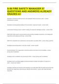
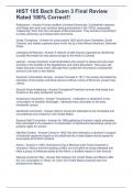
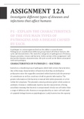
● By the fourth week, a laryngotracheal diverticulum develops from the floor of the primordial pharynx.
● The laryngotracheal diverticulum becomes separated from the foregut by tracheoesophageal folds
that fuse to form a tracheoesophageal septum. This septum results in the formation of the esophagus
and laryngotracheal tube
● The endoderm of the laryngotracheal tube gives rise to the epithelium of the lower respiratory organs
and tracheobronchial glands. The splanchnic mesenchyme surrounding the laryngotracheal tube forms
the connective tissue, cartilage, muscle, and blood and lymphatic vessels of these organs.
● Pharyngeal arch mesenchyme contributes to formation of the epiglottis and connective tissue of the
larynx. The laryngeal muscles are derived from mesenchyme in the caudal pharyngeal arches. The
laryngeal cartilages are derived from neural crest cells.
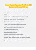
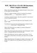
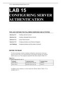
● The distal end of the laryngotracheal diverticulum forms a respiratory bud that divides into two
bronchial buds. Each bronchial bud soon enlarges to form a main bronchus, and then the main bronchus
subdivides to form lobar, segmental, and subsegmental branches
● Each tertiary bronchial bud (segmental bronchial bud), with its surrounding mesenchyme, is the
primordium of a bronchopulmonary segment. Branching continues until approximately 17 orders of
branches have formed. Additional airways are formed after birth, until approximately 24 orders of
branches are present.
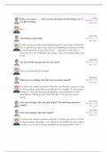
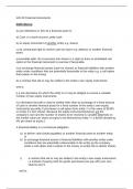
● Lung development is divided into four stages: the pseudoglandular(6–16 weeks), canalicular (16–26
weeks),terminal sac (26 weeks to birth), and alveolar (32weeks to approximately 8 years of age) stages.
● By 20 to 22 weeks, type II pneumocytes begin to secrete pulmonary surfactant. Deficiency of
surfactant results in RDS or hyaline membrane disease.
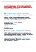
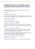
● A TEF, which results from faulty partitioning of the foregut into the esophagus and trachea, is usually
associated with esophageal atresia.
● By the fourth week, a laryngotracheal diverticulum develops from the floor of the primordial pharynx.
● The laryngotracheal diverticulum becomes separated from the foregut by tracheoesophageal folds
that fuse to form a tracheoesophageal septum. This septum results in the formation of the esophagus
and laryngotracheal tube
● The endoderm of the laryngotracheal tube gives rise to the epithelium of the lower respiratory organs
and tracheobronchial glands. The splanchnic mesenchyme surrounding the laryngotracheal tube forms
the connective tissue, cartilage, muscle, and blood and lymphatic vessels of these organs.
● Pharyngeal arch mesenchyme contributes to formation of the epiglottis and connective tissue of the
larynx. The laryngeal muscles are derived from mesenchyme in the caudal pharyngeal arches. The
laryngeal cartilages are derived from neural crest cells.
● The distal end of the laryngotracheal diverticulum forms a respiratory bud that divides into two
bronchial buds. Each bronchial bud soon enlarges to form a main bronchus, and then the main bronchus
subdivides to form lobar, segmental, and subsegmental branches
● Each tertiary bronchial bud (segmental bronchial bud), with its surrounding mesenchyme, is the
primordium of a bronchopulmonary segment. Branching continues until approximately 17 orders of
branches have formed. Additional airways are formed after birth, until approximately 24 orders of
branches are present.
● Lung development is divided into four stages: the pseudoglandular(6–16 weeks), canalicular (16–26
weeks),terminal sac (26 weeks to birth), and alveolar (32weeks to approximately 8 years of age) stages.
● By 20 to 22 weeks, type II pneumocytes begin to secrete pulmonary surfactant. Deficiency of
surfactant results in RDS or hyaline membrane disease.
● A TEF, which results from faulty partitioning of the foregut into the esophagus and trachea, is usually
associated with esophageal atresia.